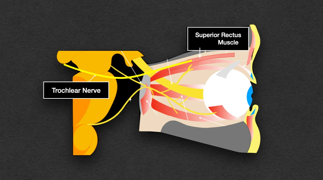
It is the fourth cranial nerve, that is purely motor and supplies only one muscle i.e., superior oblique muscle.
Characteristics
- Most slender and smallest cranial nerve.
- Only cranial nerve that emerges on the dorsal aspect of the brain.
- Only cranial nerve whose nuclear fibres decussate before emerging on the surface of the brain.
- Nucleus receives only ipsilateral corticonuclear fibres.
- Phylogenetically, it is the nerve of third eye.
Functional Components
General Somatic Efferent Fibres
- Arise from the trochlear nucleus in the midbrain.
- Supplies the superior oblique muscle of the eyeball.
Anatomy & Pathway
- Arise from the dorsal aspect of the midbrain, one on either side of frenulum veli.
- It winds round the superior cerebellar peduncle and cerebral peduncle just above the pons.
- Then, it passes between the posterior cerebral and superior cerebellar arteries, and, lies medial to and below the free margin of tentorium cerebelli.
- It enters the cavernous sinus by piercing the posterior corner of its roof.
- In the cavernous sinus, it runs forward in its lateral wall between the oculomotor and ophthalmic nerves.
- In the anterior part of cavernous sinus, it crosses over the oculomotor nerve and becomes lateral to it.
- It enters the orbit through the superior orbital fissure, superolateral to the tendinous ring.
- It runs medially above the levator palpebrae superioris to enter the orbital surface of the superior oblique which it supplies.
Clinical Significance
Trochlear nerve injury causes paralysis of the superior oblique muscle of the eyeball, that presents clinically as:
- Extorsion of the eyeball: Top of the eye twists/rotates away from the nose.
- Weakness of downward gauge: Patient faces difficulty while going downstairs or reading newspaper.
- Diplopia: Double vision when the patient looks laterally and in glances on looking downward.
References
| [](https://amzn.to/3Ixnict)
Textbook of Anatomy Head, Neck, and Brain (Volume III), Vishram Singh |
- The image used is licensed under the Creative Commons Attribution 4.0 International license. (Author: Romano, N., Federici, M. & Castaldi, A. Source: https://doi.org/10.1186/s13244-019-0719-5.)
*This article is an excerpt from the above mentioned sources and Medical Sutras does not make any ownership or affiliation claims.