It is the 5th cranial nerve, that consists of both motor and sensory fibres.
- Consists of three large nerves (ophthalmic, maxillary and mandibular), hence, the name trigeminal.
- Largest cranial nerve.
- The sensory ganglion is largest in the body and located within the cranial cavity.
Functional Components
General Somatic Afferent Fibres
- Exteroceptive sensations (Pain, touch and temperature): From the skin of head and face, mucous membrane of mouth, nasal cavity, meninges, etc. They are pseudounipolar and terminate in the main sensory nucleus and spinal nucleus of the trigeminal nerve.
- Proprioceptive sensations: From the muscles of mastication, TMJ and teeth. The neurons are unipolar and terminate in the mesencephalic nucleus of the trigeminal nerve and the reticular formation of brainstem.
Special Visceral Efferent Fibres
- Arise from the motor nucleus of the trigeminal nerve in the pons.
- Supplies the muscles derived from 1st pharyngeal arch i.e., muscles of mastication, mylohyoid, anterior belly of digastric, tensor palatini, and tensor tympani.
Anatomy & Pathway
The trigeminal nerve arises from the ventrolateral aspect of the pons at its junction with the middle cerebellar peduncle, by two roots:
- Motor root (smaller and medial).
- Sensory root (larger and lateral).
Motor Root
- Passes forward and laterally deep to the sensory root and trigeminal ganglion and enters the infratemporal fossa through foramen ovale.
- After exiting foramen ovale, it joins the mandibular nerve.
Sensory Root
-
Passes forward and laterally over the apex of the petrous temporal bone to enter the middle cranial fossa.
-
In the middle cranial fossa, it exhibits a rounded enlargement, the gasserian (trigeminal) ganglion. The ganglion occupies a dural invagination in a shallow fossa on the anterior surface of the petrous temporal bone.
-
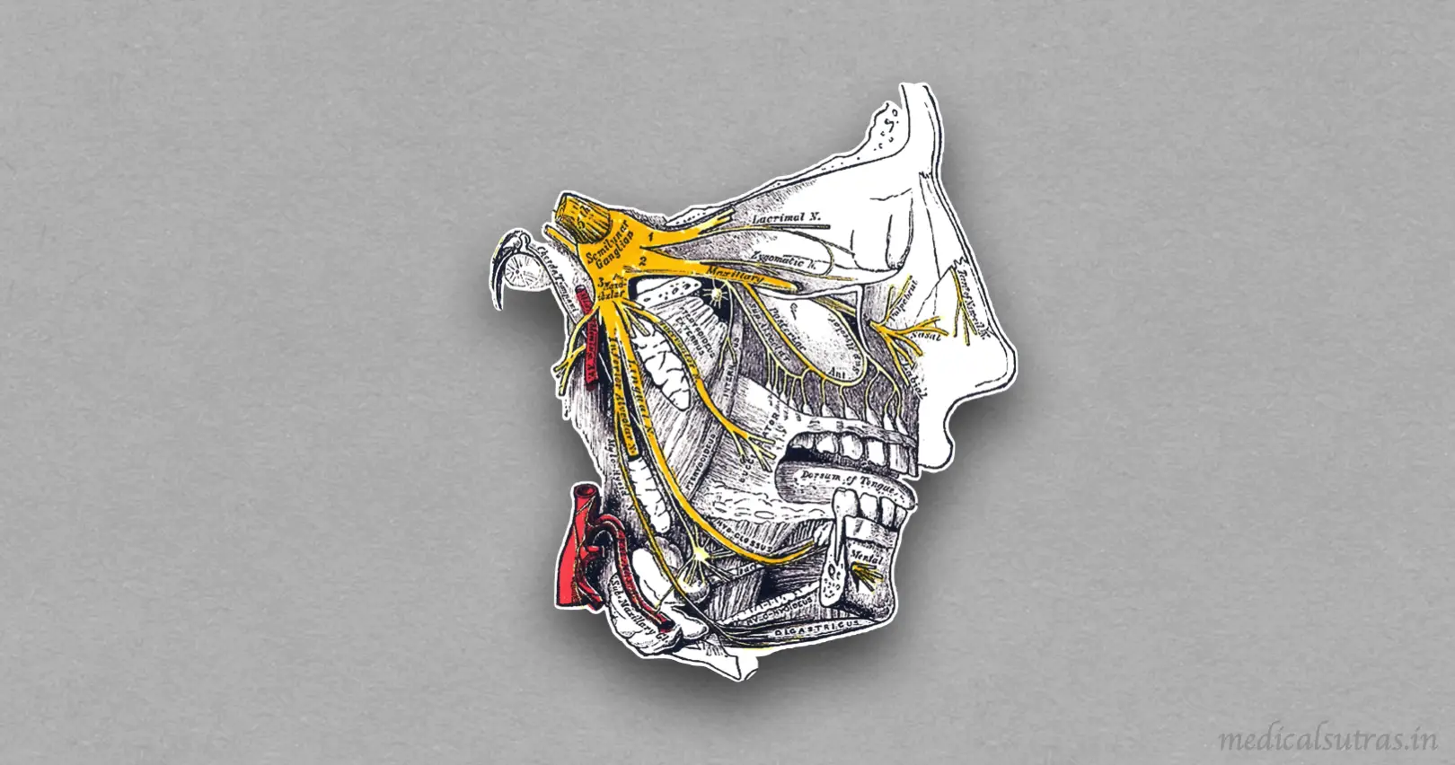
The convex distal surface of the ganglion gives origin to three large divisions of the trigeminal nerve:
- Ophthalmic nerve (sensory): Arises from the anterolateral aspect and enters the lateral wall of cavernous sinus (lies below the trochlear nerve). It divides into three branches in the cavernous sinus i.e., nasociliary, lacrimal and frontal. All these branches enter the orbit through superior orbital fissure.
- Maxillary nerve (sensory): Enters the lateral wall of cavernous sinus and occupies the lowest position. It leaves the sinus and enters the pterygopalatine fossa through the foramen rotundum.
- Mandibular nerve (mixed): Enters the infratemporal fossa through the foramen ovale.
Clinical Significance
- Trigeminal neuralgia (tic douloureux): Characterised by episodes of severe pain of sudden onset and short duration along the distribution of one or more divisions of the nerve. Maxillary and mandibular divisions are most commonly involved.
- Meckel's cave: The subarachnoid expansion/dural recess in the posteromedial portion of the middle cranial fossa that houses the Gasserian ganglion and proximal rootlets of the trigeminal nerve. It serves as a major pathway in perineural spread of pathologies such as head and neck neoplasms, and a key structure to assess in cases of trigeminal neuralgia (the sensory root can be cut here to relieve the pain of trigeminal neuralgia).
References
| [](https://amzn.to/3Ixnict)
Textbook of Anatomy Head, Neck, and Brain (Volume III), Vishram Singh |
- The image used is licensed under the Creative Commons Attribution-Share Alike 3.0 Unported license. (Author: Lipothymia, medical illustrator. Source: Wikimedia Commons.)
*This article is an excerpt from the above mentioned sources and Medical Sutras does not make any ownership or affiliation claims.