Tongue is a muscular organ situated in the floor of the mouth, associated with the functions of taste, speech, chewing, deglutition and cleansing of mouth.
Parts of Tongue
The tongue can be divided into three parts: root, tip and body.
Root
- Attached to the styloid process and soft palate above, and to mandible and hyoid bone below. These attachments prevent us from swallowing the tongue itself.
Tip
- Forms the anterior free end, that lies behind the anterior teeth.
Body The body can be divided into three parts: oral part in the anterior two-thirds, pharyngeal part in the posterior one-third and a small posteriormost part.
1. Oral/Papillary part
- It refers to the anterior two-thirds of the tongue that is placed on the floor of the mouth.
- It's superior surface is coverd with papillae, and shows a median furrow.
- The inferior surface is covered with smooth mucous membrane and shows a median fold, called frenulum linguae. On the lateral side, there is a fold called plica fimbriata that is directed forwards and medially towards the tongue tip.
2. Pharyngeal/Lymphoid part
- It refers to the posterior one-third of the tongue that lies behind the palatoglossal arches and sulcus terminalis.
- Sulcus terminalis is a faint V-shaped groove that separates the anterior and posterior parts. It forms a median pit at the centre, called foramen caecum, and runs laterally and forwards upto the palatoglossal arches.
- Foramen caecum represents the site from which the thyroid diverticulum grows down in the embryo.
- Posterior surface of the phyaryngeal part, also called the base of the tongue, forms the anterior wall of the oropharynx.
- It does not present any papillae, but have several lymphoid follicles that form the lingual tonsil.
3. Posteriormost part
- It is a small part posterior to the pharyngeal part that is connected to the epiglottis.
- It is connected to epiglottis by three folds of mucous membrane: median glosoepiglottic fold and right and left lateral glosoepiglottic folds.
- The depression on either side of median glosoepiglottic fold is called the vallecula. It is separated from the piriform fossa by the lateral folds.
Surfaces
1. Upper surface or dorsum
- It is curved and covex in all directions.
- It is divided into anterior two-thirds (oral part), posterior one-third (pharyngeal part), and small posteriormost part.
2. Inferior surface
- It is confined to the oral part only.

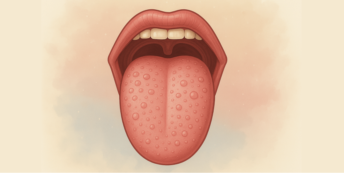
Mucous Membrane and Papillae
The mucous membrane or corium consists of projections in the anterior two-thirds. These are referred as papillae, and gives the tongue its characteristic roughness.
Types of Papillae
1. Vallate/Circumvallate papillae
- Located immediately anterior to the sulcus terminalis.
- Each papilla presents as cylindrical projection, surrounded by a circular sulcus, with taste buds on the walls.
- Large in size (Diameter: 1-2 mm).
- 8-12 in number.
2. Fungiform papillae
- Located near the tip and margins of the tonue, with some papille scattered over the dorsum.
- Each papilla consists of a narrow pedicle and a large rounded head.
- Bright red in color.
- Smaller than vallate papillae but larger than filiform.
3. Filliform/Conical papillae
- Cover the presulcal area of the dorsum of the tongue.
- Give the tongue characteristic velvety appearance.
- Each papilla is pointed and covered with keratin, with the apex often split into filamentous process.
- Smallest in size and most in number.
4. Foliate papillae
- Present at the lateral border of tongue, just anterior to circumvallate papillae.
- These are red colored and appear as short vertical folds (leaf-shaped ridges), covered with a non-keratinized epithelium.
- Around 20 in number.
- Involved in gustation, the detection of different tastes, and contain numerous taste bud.

Image Source: https://cnx.org/contents/FPtK1zmh@8.25:fEI3C8Ot@10/Preface
References
*This article is an excerpt from the above mentioned sources and Medical Sutras does not make any ownership or affiliation claims.