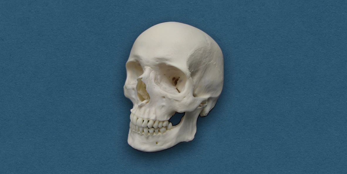
Skull refers to the skeleton of the head i.e., the entire bony structure of the head, which provides a protective case for the brain and its coverings (meninges).
Although, the terms skull and cranium are used interchangeably, cranium is a part of the skull that encloses the brain, and does not include mandible.
Bones of Skull
The skull is made up of 22 bones, that can be divided into main groups:
- Neurocranium (Cranial skeleton)
- Viscerocranium (Facial skeleton)
Neurocranium (Cranial skeleton)
- It is the upper part of the skull that is rigid and strong.
- It can further be subdivided into an upper dome-shaped part called the calvaria, cranial vault or brain box, and a lower part referred as the skull base.
| **Paired Bones (2)** | **Unpaired Bones (4)** |
|---|---|
| - Parietal - Temporal | - Frontal - Occipital - Sphenoid - Ethmoid |
*Malleus, Incus, and Stapes are also described as paired bones that forms part of the neurocranium.
Viscerocranium (Facial skeleton)
- It refers to lower anterior part of skull, that is rather fragile and forms the basis of face.
- It forms the rest of the skull and includes the mandible.
| **Paired Bones (6)** | **Unpaired Bones (2)** |
|---|---|
| - Maxilla - Zygomatic - Nasal - Lacrimal - Palatine - Inferior nasal concha | - Mandible - Vomer |
Joints of Skull
Most of the bones of the skull are joined by sutures, a few primary cartilaginous joints and synovial joints.
Sutures
- Coronal suture (serrate): Frontal and parietal bones.
- Parietotemporal suture (squamous): Parietal and temporal bones.
- Lambdoid suture (denticulate): Parietal and occipital bones.
- Internasal suture (plane): Between the nasal bones.
Synovial joints
- Temporomandibular joint, that joins the mandible to the cranium (temporal bone).
- Atlanto-occipital joint, that joins the base of skull (occipital bone) with the vertebral column (C1 vertebra - Atlas).
Primary cartilaginous joints (synchondrosis)
- Spheno-occipital synchondrosis.
Anatomical Position of Skull
In the anatomical position, the orbital cavities of the skull are directed forwards. It can be placed in this position along the following two planes:
- Frankfurt's Horizontal Plane: It is formed by a horizontal line joining the infraorbital margin and the upper margin of external auditory meatus.
- Reid's Base Line: It is formed by a horizontal line passing through the infraorbital margin and the center of external auditory meatus.
Methods of Studying Skull
The skull must be studied as a whole along with individual bones, to gain a clear understanding. The different methods include:
-
Study from the outside (Exterior of the skull)
- Superior view (Norma verticalis)
- Posterior view (Norma occipitalis)
- Anterior view (Norma frontalis)
- Lateral view (Norma lateralis)
- Inferior view (Norma basalis)
-
Study from the inside (Interior of the skull)
- Internal surface of the cranial vault
- Internal surface of the cranial base
-
Study of individual bones.
Additional Points
Fontanelles
These are unossified areas between the bones of the skull, that permits growth of the brain and some overlapping of skull bones (moulding) during childbirth.
These are 6 fontanelles at birth, at the four angles of parietal bones.
- Anterior fontanelle (1): It is diamond-shaped, largest fontanelle, located at the junction of parietal and frontal bones. It closes by 2 years of age.
- Posterior fontanelle (1): It is triangular-shaped, situated at the junction of sagittal and lambdoid sutures. It closes soon after birth.
- Lateral fontanelles (4): These include sphenoidal (anterolateral) fontanelle and mastoid (posterolateral) fontanelle, located at the sphenoid and mastoid angles of parietal bones. These close within a few weeks after birth.
*Clinical significance
- The degree of tenseness of the anterior fontanelle membrane gives an index of intracranial pressure. An abnormal depression indicates dehydration (insufficiency of body fluids).
- Anterior fontanelle lies just underneath the superior sagittal sinus, and allows access to superior sagittal sinus, and needle can be passed into the lateral ventricle of brain through its lateral angle.
Pneumatic Bones
Refers to the bones that have air cells in them, for example, frontal and maxillary bone.
- Reduce the weight of the skull.
- Maintain humidity of inspired air.
- Give resonance to voice.
- May get infected, leading to sinusitis.
Emissary Veins
These veins connect the intracranial venous sinuses with extracranial veins.
- They help to relieve raised intracranial pressure.
- However, they may spread infection to the cranial venous sinuses, since, they are valveless.
| **Emissary Vein** | **Venous Sinus** | **External Vein** |
|---|---|---|
| 1\. Parietal emissary vein
(Parietal foramen) | Superior sagittal sinus | Veins of scalp |
| 2\. Mastoid emissary vein
(Mastoid foramen) | Sigmoid sinus | Veins of scalp |
| 3\. Emissary vein
(Hypoglossal canal) | Sigmoid sinus | Internal jugular vein |
| 4\. Condylar emissary vein
(Posterior condylar foramen) | Sigmoid sinus | Suboccipital venous plexus |
| 5\. Emissary veins
(Foramen lacerum) | Cavernous sinus | Pharyngeal venous plexus |
| 6\. Emissary vein
(Foramen ovale) | Cavernous sinus | Pterygoid venous plexus |
| 7\. Emissary vein
(Foramen caecum) | Superior sagittal sinus | Veins from upper part of nose |
References
- BD Chaurasia's Human Anatomy Volume 3 (Head & Neck), Eighth edition, CBS Publishers and Distributors Pvt Ltd
- Image Source: Wikimedia Commons. Author: Sklmsta. This file is made available under the Creative Commons CC0 1.0 Universal Public Domain Dedication.
*This article is an excerpt from the above mentioned sources and Medical Sutras does not make any ownership or affiliation claims.