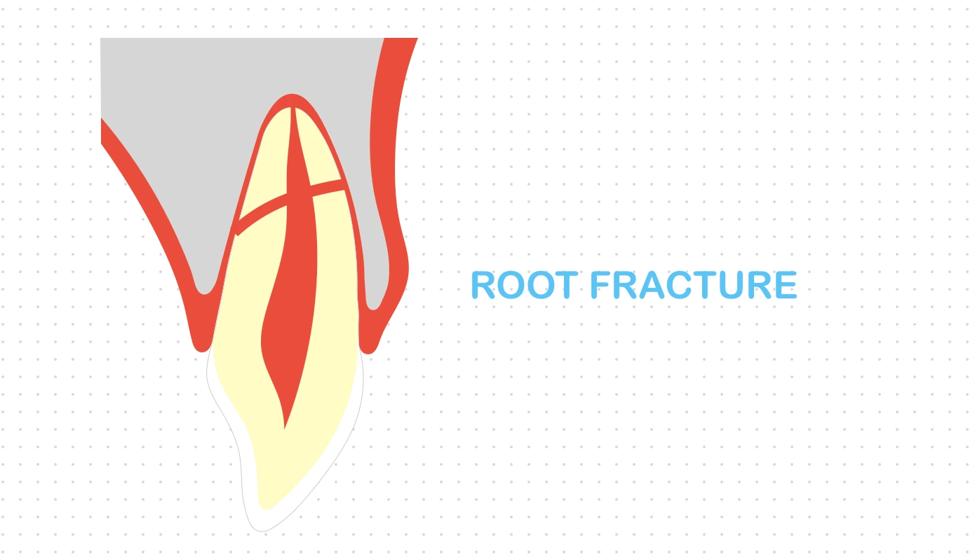
Root fracture involves fracture of the dentin, pulp and cementum, usually caused by a frontal impact which creates compression zones labially and lingually.
The fracture may be horizontal, oblique or a combination of both, depending on the shearing stress zone.
Clinical Findings
- The coronal segment may be mobile, slightly extruded and displaced in an oral direction.
- The tooth may be tender on percussion.
- Bleeding from the gingival sulcus may be seen.
- Pulp sensibility testing may be negative initially, indicating transient or permanent neural damage.
Radiographic Findings
-
While the fracture may be located at any level of the root, the fracture line is mostly oblique and at an optimal angle for radiographic disclosure.
-
Normally visible when the central beam is directed within a maximum range of 15-20o of the fracture plane.
-
Ellipsoid radiolucent line: Two additional radiographs should be taken
- At increased angulation of 15o to the original.
- At negative angulation of 15o to the original.
-
Occasionally, root fractures may go undetected initially, but found in later radiographs due to
- Development of haemorrhage or granulation tissue between the fragments, which displaces the coronal fragment incisally, or,
- Resorption at the fracture line.
-
CBCT can be considered to determine the location, extent and direction of the fracture.
Root Resorption & Fracture Healing
Healing events following root fracture are initiated at the site of pulp and PDL involvement, and depend on the pulp status (intact, severely stretched or infected with bacteria) and stage of root development.
The resorption process is usually detected within 1 year after injury and precedes fracture healing and obliteration of the pulp canal.
Root Resorption
- External surface resorption (ESR) characterised by rounding of the fracture edges medially and/or distally, at the periodontal side of the fracture.
- Internal surface resorption (ISR) characterised by rounding of the fracture edges centrally, at the pulpal side of the fracture, significantly related to hard tissue union.
- Internal tunneling resorption (ITR), burrows behind the predentin layer and along the root canal walls of the coronal fragment.
Healing with Calcified Tissue
- Seen in cases where the pulp is intact (reacts similar to a coronal pulp exposure with intact vasculature and no infection) with little or no dislocation (i.e, concussion or subluxation) of the coronal fragment.
- There is formation of a uniting callus of hard tissue at the fracture line and partial pulp canal obliteration, confined to the apical fragment
- In most cases, the innermost layer of repair seems to be dentin, while the more peripheral part of the fracture is incompletely repaired with cementum.
- Radiographically, a fracture line is often discernible, and clinically, the tooth shows normal mobility with normal reaction to percussion and normal or slightly decreased response to pulpal sensibility testing.
Interposition of Connective Tissue (PDL)
- Seen in cases where the pulp is severed or severely stretched (moderate pulpal injury such as extrusion or lateral luxation of the coronal fragment) but not contaminated with bacteria.
- There is revascularisation of the coronal pulp, while the periodontally derived cells dominate root fracture healing, resulting on the union of the coronal and apical root fragments by interposition of connective tissue.
- Histologically, this is characterised by the presence of cementum covering the fracture surfaces and connective tissue between the fragments.
- Radiographically, peripheral rounding of the fracture edges (external surface resorption) and radiolucent line separating the fragments can be seen.
- Clinically, the tooth shows firm or slight mobility, with weak pain response to percussion and normal pulp tests.
Interposition of Bone & Connective Tissue (PDL)
- Seen in cases of trauma prior to completed growth of the alveolar process, wherein the coronal fragment continues to erupt, while the apical fragment remains stationary in the jaw.
- There is interposition of a bony bridge and connective tissue between the apical and coronal fragments with normal PDL surrounding both fragments.
- Radiographically, a bony bridge separating the fragments and periodontal space around both fragments is seen, along with complete pulp canal obliteration in both fragments.
- Clinically, the tooth is firm and react normally to pulp tests.
Interposition of Granulation Tissue
- Seen in cases of severely stretched pulp that gets contaminated with bacteria, resulting in infection and necrosis of the coronal pulp.
- There is accumulation of inflamed, granulation tissue between the two root fragments.
- Radiographically, widening of the fracture line, loss of lamina dura and rarefaction of the alveolar bone corresponding to the fracture line is seen.
- If the tooth is not splinted, the coronal fragment is loose, slightly extruded and sensitive to percussion. If splinted, the apical fragment becomes displaced in apical direction.
Management
The treatment protocol involves reduction of displaced coronal fragments and immobilisation.
- The coronal fragment should be repositioned immediately by digital manipulation. If any resistance is felt while repositioning, the alveolar bone should be evaluated for any fracture and repositioned.
- The position of the root should be checked radiographically after repositioning.
- The mobile coronal fragment should be stabilised with a firm and rigid splint that is passively applied (eg. acid-etch resin splint) for 4 weeks. If the fracture is located cervically, stabilisation for a longer period of time (upto 4 months) may be needed.
- No endodontic treatment should be started at the emergency visit.
- It is recommended to monitor healing of the fracture for at least one year. Pulp status should also be monitored.
In mature teeth where the cervical fracture line is located above the alveolar crest and the coronal fragment is very mobile
- Remove the coronal fragment, followed by root canal treatment and restoration with a post-retained crown.
- Additional treatment options such as orthodontic or surgical extrusion of the apical segment, crown lengthening surgery, or even extraction may be considered in future (similar to those for crown-root fractures).
Endodontic Considerations
Pulp Necrosis
- Usually occurs in the coronal fragment only, the apical segment rarely undergoes pathological changes that require treatment.
- Only the coronal segment may require endodontic treatment.
Root Resorption
- External surface resorption, Internal surface resorption and Internal tunneling resorption are all self-limiting and resolves within the first 1-2 years after injury. They don't require any interceptive treatment, however, needs to be monitored and distinguished from bone resorption at the level of root fracture which is indicative of pulp necrosis.
- External inflammatory resorption: Needs to be treated with pulp removal and root filling.
- External replacement resorption (Ankylosis): Cannot be treated.
Prognosis
Clinical and radiographic evaluations (follow up) are necessary at 4 weeks (splint removal for mid-third and apical third fractures ), 6-8 weeks, 4 months (splint removal for cervical third fractures), 6 months, 1 year and then yearly for at least 5 years.
Favourable Outcomes
- Positive response to pulp sensibility testing, however, a false negative response is possible for several months. (Endodontic treatment should not be started solely on the basis of no response to pulp sensibility testing.)
- Signs of repair between the fractured segments.
- Normal or slightly more than physiologic mobility of the coronal fragment.
Unfavourable Outcomes
- Symptomatic
- Extrusion and/or excessive mobility of the coronal segment.
- Radiolucency at the fracture line.
- Pulp necrosis and infection with inflammation in the fracture line.
Deciduous Dentition
If the coronal fragment is not displaced, no treatment is required. Follow up after 1 week, 6-8 weeks, 1 year and, then, yearly follow up until eruption of permanent teeth.
If the coronal fragment is displaced and,
-
Not excessively mobile: Leave the coronal fragment to spontaneously reposition, even if there is some occlusal interference.
-
Excessively mobile and interfering with occlusion:
- Extract the loose coronal fragment, and leave the apical fragment in place for resorption.
- Reposition the loose coronal fragment gently. If it is unstable, stabilise with a flexible splint for 4 weeks.
-
Follow up after 1 week, 4 weeks for splint removal, 8 weeks, 1 year and, then, yearly follow up until eruption of permanent teeth.
Parent/Patient Education
- Care should be taken while eating, so as not to further traumatise the injured tooth.
- The affected area should be cleaned with a soft brush or clean cotton swab, along with alcohol-free 0.1-0.2% Chlorhexidine mouth rinse applied topically twice a day for 1 week. This is to prevent plaque accumulation and proper gingival healing.
Points to Note
- Root fracture in immature teeth with incomplete root formation, may present as a partial fracture seen as a unilateral break in the continuity of the root surface. (Similar to green stick fractures of long bones.)
- Rigid fixation such as orthodontic bands are contraindicated for splinting (immobilisation) : A passively applied splint is recommended because active forces may further injure the already traumatised pulp and lead to pulp necrosis.
- Immature teeth with incomplete root fractures require no fixation and will heal by hard tissue union.
- It has been found that negative pulp sensitivity tests at the time of injury are significantly related to later pulp necrosis, presumably a reflection of severe pulp injury.
- In case of fracture located at the cervical third of the root, the coronal fragment, esp. if not mobile, should not be removed at the emergency visit, since cervical fractures have the potential to heal.
- In cases where extraction is required, the marginal socket wall should be preserved, otherwise, it can lead to collapse of the alveolar process.
References
- Textbook and Color Atlas of Traumatic Injuries to the Teeth, Jens O. Andreasen, Frances M. Andreasen, Lars Andersson. https://amzn.to/4bd3iIg
- Bourguignon C, Cohenca N, Lauridsen E, et al. International Association of Dental Traumatology guidelines for the management of traumatic dental injuries: 1. Fractures and luxations. Dent Traumatol. 2020;36:314–330. https://doi.org/10.1111/edt.12578.
- Day P, Flores MT, O'Connell A, et al. International Association of Dental Traumatology guidelines for the management of traumatic dental injuries: 3. Injuries in the primary dentition. Dent Traumatol. 2020;36:343–359. https://doi.org/10.1111/edt.12576
*This article is an excerpt from the above mentioned sources and Medical Sutras does not make any ownership or affiliation claims.