The primary maxillary second molar is similar in characteristic to the permanent maxillary first molar, but, is smaller.
Chronology
- First evidence of calcification: 19 weeks in utero.
- Crown completion: 11 months.
- Eruption: 29 months.
- Root completion: 3 years.
Dimensions
- Overall length: 17.5 mm
- Crown length: 5.7 mm
- Root length: 11.7 mm
- Mesiodistal diameter of crown: 8.2 mm
- Mesiodistal diameter of crown at cervix: 6.4 mm
- Labiolingual diameter of crown: 10.0 mm
- Labiolingual diameter of crown at cervix: 8.3 mm
Crown Morphology
Buccal Aspect
- Crown is much larger than the primary first molar.
- Shows two well-defined buccal cusps (nearly equal in size), with a buccal developmental groove between them.
- Crown is narrow at the cervix in comparison to MD measurement at the contact areas.
Lingual Aspect
-
Shows three cusps: two well developed mesiolingual and distolingual cusps, and a third poorly developed supplemental cusp known as Tubercle of Carabelli, or the fifth cusp.
- It lies apical to the ML cusp and acts as a buttress or supplement to the bulk of the ML cusp.
- If the fifth cusp is missing, some traces of developmental lines or dimples remain.
-
The ML and DL cusps are separated by a well-defined developmental groove, which connects with the developmental groove outlining the fifth cusp.
Mesial Aspect
- Crown has typical molar outline, resembling permanent molars.
- Crown appears short as its buccolingual width is shorter in comparison to its length.
- Mesiolingual cusp with its fifth cusp appears large in comparison to the mesiobuccal cusp.
- Mesiobuccal cusp appears relatively short and sharp, and is directly below the bifurcation of MB and lingual root.
Distal Aspect
- The distal measurement of the crown is less than the mesial measurement.
- The lingual outline is smooth and rounded, while, the buccal surface is almost straight from the crest of curvature to the tip of the buccal cusp.
- Distobuccal and distolingual cusps are about the same length.
- Cervical line is approximately straight, as found mesially.
Occlusal Aspect
- The tooth resembles permanent maxillary first molar.
- Shape is somewhat rhomboidal, with four well-developed cusps and one supplemental cusp.
- Buccal surface is flat with developmental groove between the cusps (less prominent compared to permanent first molar).
- Occlusal surface has a central fossa with a central pit, well defined mesial triangular fossa, distal to the mesial marginal ridge with a mesial pit at its center.
- Central groove is well-defined, present at the bottom of a sulcus, connecting mesial triangular fossa with the central fossa.
- Buccal developmental groove extends buccally from the central pit, separating the triangular ridges (occlusal continuations of MB and DB cusps).
- Oblique ridge is prominent and connects the ML cusp with the DB cusp.
- Distal fossa lies distal to the oblique ridge and harbors the distal developmental groove.
- Distal groove acts a line of demarcation between the ML and DL cusps, and continues on to the lingual surface as lingual developmental groove.
- Distal marginal ridge is as well developed as the mesial marginal ridge. (Marginal ridges are not developed equally in the primary maxillary first molars.)

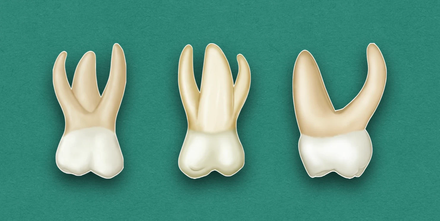
Root Morphology
Buccal Aspect
- Root appears slender.
- Longer and heavier than the roots of primary first molar.
- Point of bifurcation between the buccal roots is close to the cervical line of the crown.
Lingual Aspect
- All three roots can be seen.
- Lingual root is large and thick compared to other two roots, however, it is approximately the same length as the MB root.
Mesial Aspect
- Mesiobuccal root appears broad and flat, and extends lingually far out beyond the crown outline.
- Lingual root have somewhat similar curvature as lingual root of primary maxillary first molar.
- Point of bifurcation between MB and lingual root is 2-3 mm apical to the cervical line.
Distal Aspect
- All three are seen, although the distobuccal root is superimposed on the mesiobuccal root.
- Distobuccal root is shorter and narrower than the other roots.
- Point of bifurcation between DB and lingual root is more apical than any other points of bifurcation, and is nearly centered above the crown.
References
- Wheeler's Dental Anatomy, Physiology and Occlusion(2019), Stanley J. Nelson DDS MS, Elsevier.
*This article is excerpt from the above mentioned book and Medical Sutras does not make any ownership and affiliation claims.