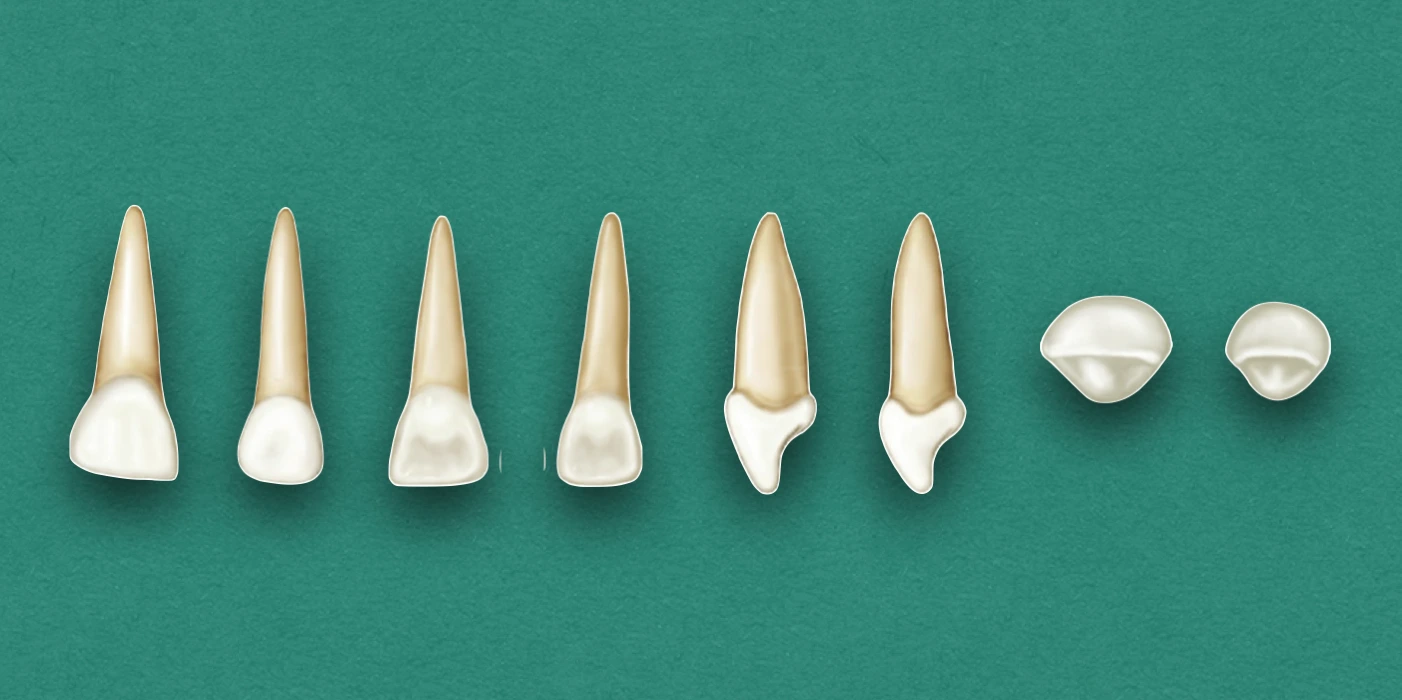
Generally, the primary maxillary central and lateral incisors are similar in morphology, and the key difference is in dimensions of the two teeth.
Points differentiating Lateral Incisor from Central Incisor:
- Smaller crown size in all directions.
- Cervicoincisal length is greater than mesiodistal width.
- Distoincisal angles are more rounded.
- The ratio of root length to crown length is more i.e., root is longer in proportion to crown.
Chronology
Primary Maxillary Central Incisor
- First evidence of calcification: 14 weeks in utero.
- Crown completion: 1 and half months.
- Eruption: 10 months.
- Root completion: 1 and half years.
Primary Maxillary Lateral Incisor
- First evidence of calcification: 16 weeks in utero.
- Crown completion: 2 and half months.
- Eruption: 11 months.
- Root completion: 2 years.
Dimensions
Primary Maxillary Central Incisor
- Overall length: 16.0 mm
- Crown length: 6.0 mm
- Root length: 10.0 mm
- Mesiodistal diameter of crown: 6.5 mm
- Mesiodistal diameter of crown at cervix: 4.5 mm
- Labiolingual diameter of crown: 5.0 mm
- Labiolingual diameter of crown at cervix: 4.0 mm
Primary Maxillary Lateral Incisor
- Overall length: 15.8
- Crown length: 5.6
- Root length: 11.4
- Mesiodistal diameter of crown: 5.1
- Mesiodistal diameter of crown at cervix: 3.7
- Labiolingual diameter of crown: 4.0
- Labiolingual diameter of crown at cervix: 3.7
Crown Morphology
Labial Aspect
- Mesiodistal diameter is greater than cervicoincisal length (opposite in case of primary lateral and permanent central incisors).
- Labial surface is smooth.
- Incisal edge is almost straight.
- Developmental lines not usually seen.
Lingual Aspect
- Well-developed marginal ridges.
- Highly developed cingulum, extending up toward the incisal ridge and partially dividing the concave lingual surface below the incisal edge into a mesial and distal fossa.
Mesial & Distal Aspects
- Crown appears wider in relation to its total length, when measured at the cervical third.
- Crown appears thick in the middle and even down toward the incisal third.
- The curvature of the cervical line (cementoenamel junction) is distinct, and curves toward the incisal ridge.
- Cervical curvature is less on the distal side than the mesial side (similar to permanent central incisor).
Incisal Aspect
- Incisal edge is relatively straight, and is centered over the main bulk of the crown.
- Labial surface appears much broader and smoother than lingual surface.
- Lingual surface tapers toward the cingulum.
- Mesial and distal surfaces are relatively broad towards the incisal ridge or at incisal third.
Root Morphology
Labial Aspect
- Cone-shaped with even, tapered sides.
- Root length greater than crown length (in comparison to permanent central incisor).
Lingual Aspect
- Root narrows from labial to lingual aspect and presents a ridge along its full length.
- Cross section at the crown-root junction shows somewhat triangular shape with sides formed by the labial, mesial and distal surfaces.
Mesial & Distal Aspects
- Root appears long cone shaped and even tapered, but, is more blunt compared to labial and lingual aspects.
- Medial surface have developmental groove or concavity, while distal surface is generally convex.
References
- Wheeler's Dental Anatomy, Physiology and Occlusion(2019), Stanley J. Nelson DDS MS, Elsevier.
*This article is excerpt from the above mentioned book and Medical Sutras does not make any ownership and affiliation claims.