The primary maxillary first molar is designed to be the premolar section of the primary dentition. In function it acts as a compromise between the size and shape of primary anterior teeth and the molar area.
Chronology
- First evidence of calcification: 15 and half weeks in utero.
- Crown completion: 6 months.
- Eruption: 16 months.
- Root completion: 2 and half years.
Dimensions
- Overall length: 15.2 mm
- Crown length: 5.1 mm
- Root length: 10.0 mm
- Mesiodistal diameter of crown: 7.3 mm
- Mesiodistal diameter of crown at cervix: 5.2 mm
- Labiolingual diameter of crown: 8.5 mm
- Labiolingual diameter of crown at cervix: 6.9 mm
Crown Morphology
Buccal Aspect
- The crown is widest at the contact areas mesially and distally, and it converges toward the cervix (width at the cervix is 2 mm less than that at the contact area). This results in a narrower look to the cervical portion of the crown and root, as compared to the permanent maxillary first molar.
- Occlusal line is slightly scalloped but with no definite cusp form.
- Buccal surface is smooth, with little evidence of developmental grooves.
- From buccal aspect, it appears smaller in all measurements as compared to second molar.
Lingual Aspect
- General outline is similar to the buccal aspect.
- Crown converges considerably in lingual direction, which makes the lingual portion calibrate less mesiodistally than the buccal side.
- Mesiolingual cusp is the most prominent cusp (longest and sharpest).
- Distolingual cusp is poorly defined (small and rounded when it exists at all).
- Distobuccal cusp is longer and better developed, and can be seen from the lingual aspect.
- Three-cusped molar: Presents one large lingual cusp with no developmental groove lingually.
Mesial Aspect
- Dimension at the cervical third is greater than the dimension at the occlusal third (more pronounced on primary than permanent teeth).
- Mesiolingual cusp is longer and sharper than the mesiobuccal cusp.
- Buccal outline of the cervical third presents a pronounced convexity (outstanding characteristic of this tooth).
- Cervical line shows some curvature in the direction of the occlusal surface.
Distal Aspect
- The crown tapers markedly toward the distal end, and is narrower distally.
- Distobuccal cusp is long and sharp, while the distolingual cusp is poorly developed.
- The prominent bulge seen from mesial aspect does not continue distally.
- Cervical line may curve occlusally, or extend straight across from buccal surface to lingual surface.
Occlusal Aspect
- Crown converges lingually (distance between MB and DB line angles is greater than the distance between ML and DL line angles).
- Crown converges distally (distance between MB and ML line angles is greater than than the distance between DB and DL line angles).
- The occlusal surface has a central fossa.
- Mesial triangular fossa: It is just inside the mesial marginal ridge, with a mesial pit and a sulcus with its central groove connecting the two fossae.
- Buccal developmental groove: It is well defined and divides the MB cusp and DB cusp.
- Supplemental grooves: Radiate from the pit in mesial triangular fossa: one buccally, one lingually, and one toward the marginal ridge.
- Oblique ridge: It refers to well defined triangular ridge that connects ML cusp with the DB cusp. In some teeth, the ridge is indefinite, and the central developmental groove extends from the mesial pit to the distal developmental groove.
- Distal marginal ridge: It is thin and poorly developed as compared with mesial marginal ridge.

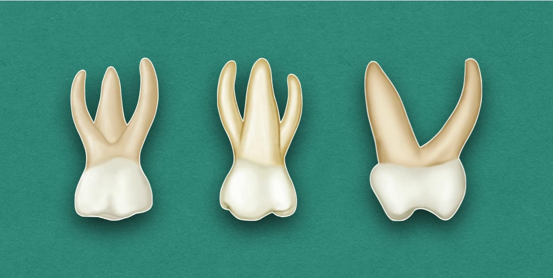
Root Morphology
- The roots are slender, long, and spread widely.
- The distal root is considerably smaller than the mesial one.
- Bifurcation of the roots begins almost immediately at the site of cervical line (CEJ). This arrangement is in effect for the entire root trunk, resulting in a trifurcation (characteristic of all primary molars). In permanent molars, the root trunk is much heavier, and there is a greater distance between the cervical lines to the points of bifurcations.
- From the mesial aspect, the lingual root appears longs and slender, and extends lingually to a marked degree. It curves sharply in buccal direction above the middle third.
References
- Wheeler's Dental Anatomy, Physiology and Occlusion(2019), Stanley J. Nelson DDS MS, Elsevier.
*This article is excerpt from the above mentioned book and Medical Sutras does not make any ownership and affiliation claims.