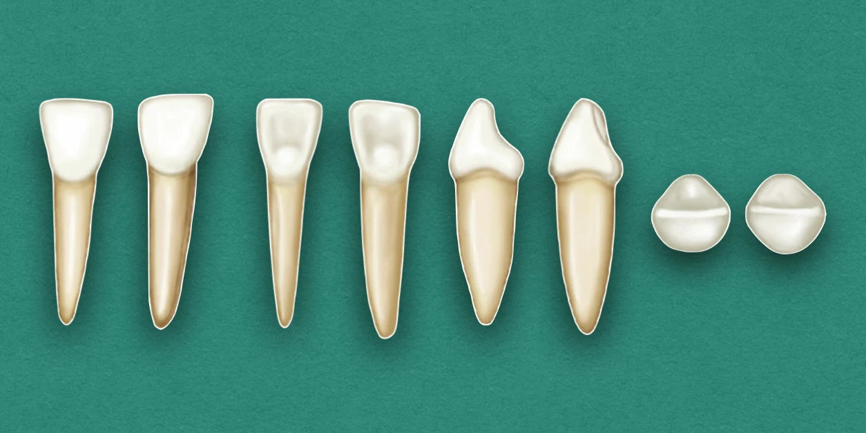
The primary mandibular central and lateral incisors are similar in outline, and supplement each other in function. They seem to be built for strenuous service.
Key differences between the two are:
- Lateral incisor is somewhat larger in all measurements except labiolingually, where the two teeth are practically identical.
- Cingulum of lateral incisor may be little more generous than that of central incisor.
- Lingual surface of crown between marginal ridges may be more concave.
- The incisal ridge slopes downward distally, resulting in lowering the distal contact area apically, so that proper contact can be made with the mesial surface of the primary mandibular canine.
Chronology
Mandibular Central Incisor
- First evidence of calcification: 14 weeks in utero.
- Crown completion: 2 and half months.
- Eruption: 8 months.
- Root completion: 1 and half years.
Mandibular Lateral Incisor
- First evidence of calcification: 16 weeks in utero.
- Crown completion: 3 months.
- Eruption: 13 months.
- Root completion: 1 and half months.
Dimensions
Mandibular Central Incisor
- Overall length: 14.0 mm
- Crown length: 5.0 mm
- Root length: 9.0 mm
- Mesiodistal diameter of crown: 4.2 mm
- Mesiodistal diameter of crown at cervix: 3.0 mm
- Labiolingual diameter of crown: 4.0
- Labiolingual diameter of crown at cervix: 3.5
Mandibular Lateral Incisor
- Overall length: 15.0 mm
- Crown length: 5.2 mm
- Root length: 10.0 mm
- Mesiodistal diameter of crown: 4.1 mm
- Mesiodistal diameter of crown at cervix: 3.0 mm
- Labiolingual diameter of crown: 4.0 mm
- Labiolingual diameter of crown at cervix: 3.5 mm
Crown Morphology
Labial Aspect
- Labially, crown appears flat with no developmental grooves.
- Mesial and distal sides are tapered evenly from the contact areas, with the measurement being less at the cervix.
- The crown is wide in proportion to its length, as compared to the permanent central incisor.
Lingual Aspect
- Presents marginal ridges and cingulum.
- May have a flattened surface level with the marginal ridges, or may present slight concavity, called lingual fossa, at the middle third and incisal third.
Mesial Aspect
- Shows the typical outline of an incisor tooth, although measurements are small.
- Incisal ridge is centered over the center of the root, and between the crest of curvature of crown, labially and lingually.
- The cervical contours are more pronounced in comparison to the prominences found on permanent central incisor.
Distal Aspect
- The outline is reverse of that found from the mesial aspect, little differences can be noted.
- Cervical line is less curved toward the incisal ridge, as compared to the mesial surface.
Incisal Aspect
- Presents a straight incisal ridge, that bisects the crown labiolingually.
- Shoes the crests of contour at the cervical third labially and lingually.
- Labial surface presents a flat surface (slightly convex), while the lingual surface is flat and slightly concave.
- On the lingual side, a definite taper is evident toward the cingulum.
Root Morphology
Labial Aspect
- Root is almost twice the length of the crown.
- It is long and evenly tapered down to the apex, which is pointed.
- The root trunk appears heavy, making the tooth resemble permanent maxillary lateral incisor.
Mesial Aspect
- Root is almost flat and evenly tapered.
- The apex presents a more blunt appearance than found with the lingual or labial aspects.
Distal Aspect
- A developmental depression is often evident on the root.
References
- Wheeler's Dental Anatomy, Physiology and Occlusion(2019), Stanley J. Nelson DDS MS, Elsevier.
*This article is excerpt from the above mentioned book and Medical Sutras does not make any ownership and affiliation claims.