Canine is the third tooth from the midline, placed at the corners of the mouth (hence, referred as cornerstones of the dental arches).
The teeth are called "canines" because of the close resemblance of the shape, location and the extra anchorage furnished by the long, strongly developed roots of canines to those of the carnivore.
Chronology
- First evidence of calcification: 4-5 months.
- Crown completion: 6-7 years.
- Eruption: 11-12 years.
- Root completion: 13-15 years.
Dimensions
- Overall length: 27.0 mm.
- Crown length: 10.0 mm.
- Root length: 17.0 mm.
- Mesiodistal diameter of crown: 7.5 mm.
- Mesiodistal diameter of crown at cervix: 5.5 mm.
- Labiolingual diameter of crown: 8.0 mm.
- Labiolingual diameter of crown at cervix: 7.0 mm.
- Cervical line curvature (Mesial): 2.5 mm.
- Cervical line curvature (Distal): 1.5 mm.
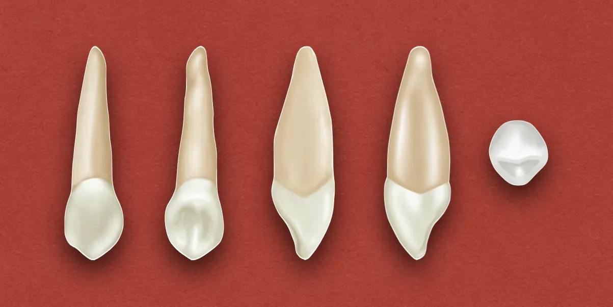
Crown Morphology
Labial Aspect
- Surface: The labial surface of crown is smooth, with no noticeable developmental lines.
- It is divided into three labial lobes by shallow depressions present mesially and distally. This produces a ridge on the labial surface, and all areas mesial to the ridge crest exhibit convexity, while, distal areas exhibit a tendency towards concavity at the cervical third of the crown.
- A line drawn over the crest of labial ridge, from cervical line to the cusp tip, is curved with mesial inclination at its center.
- Mesial outline: May be convex from the cervix to the center of mesial contact area, or may exhibit a slight concavity above the contact area.
- Distal outline: It is usually concave between the cervical line and the distal contact area.
- Contact area: Mesially, the center of contact area is approximately at the junction of middle and incisal thirds of the crown, while, the distal contact area is usually at the center of the middle third of the crown.
- Cusp: The cusp has a mesial (shorter) and a distal slope, both of which show a tendency toward concavity, before the crown is worn out.
- The cusp tip is on line with the center of the root, unless the crown is worn unevenly.
Lingual Aspect
- The crown is narrower lingually than labially.
- Cervical line: The curvature of cervical line is more even as compared to that found labially. It may be straight for a short interval.
- Cingulum: It is large and, in some cases, it is pointed, like a small cusp. In this type, definite ridges are found below the cingulum and between strongly developed marginal ridges.
- Lingual ridge: It is seen occasionally as a well-developed ridge, that is confluent with the cusp tip and extends to a point near the cingulum.
- Lingual fossae: Mesial and distal lingual fossae are evident as shallow concavities between the lingual ridge and marginal ridges.
- Sometimes, the lingual surface is smooth with confluent cingulum, marginal ridges, and lingual portion of incisal ridges, and little evidence of developmental grooves.
Mesial Aspect
- The crown is wedge-shaped, with the wedge point represented by the cusp tip.
- It generally shows greater bulk and greater labiolingual measurement than other anterior teeth.
- The mesial surface presents convexities at all points except for a small circumscribed area above the contact area where the surface is concave and flat between that area and the cervical line.
- The crest of curvature of the crown is at a level more incisal, as the middle labial and lingual lobes are more highly developed.
- Labial outline: Shows a flattened area at the cervical third, and is slightly convex from the crest of curvature at the cervical third to the cusp tip. The entire outline from cervical line to the cusp tip is more convex than the maxillary central incisor.
- Lingual outline: Shows a convex line along the cingulum, which straightens out in the middle third, and becomes convex again in the incisal third.
- Cervical line: Curves toward the cusp, approximately 2.5mm at the cementoenamel junction.
Distal Aspect
It is somewhat similar to the mesial aspect, with the following variations:
- More concave surface, usually above the contact area.
- Cervical line exhibits less curvature towards the cusp ridge.
- Distal marginal ridge is heavier and more irregular in outline.
Incisal Aspect
- The tip of the cusp is labial to the center of crown labiolingually, and mesial to the center mesiodistally. The long of axis of the root is directly in the line of vision.
- The labiolingual dimension is greater than the mesiodistal.
- Labially, the ridge of middle labial lobe is very noticeable. It attains greatest convexity at the cervical third, becoming broader and flatter at middle and incisal thirds.
- Lingually, the cingulum makes up the cervical third of the crown.
- A mesiodistal line bisecting the cusp and cusp ridges is almost always straight and bisects the short arcs of mesial and distal contact areas.
Root Morphology
Labial Aspect
- The labial surface of root is smooth and convex at all points. It appears slender as compared with the bulk of crown.
- It is conical in form with a bluntly pointed apex.
- The root commonly have a sharp curvature (mostly in mesial direction), near the apical third.
Lingual Aspect
- The lingual portion is narrower than the labial portion, and the mesial and distal root surfaces are visible from the lingual aspect.
- Mesial and distal developmental depressions can be seen on most of the roots, extending most of the root length.
- The lingual ridge is rather narrow, but, is smooth and convex at all points from the cervical line to the apical end.
Mesial Aspect
- The root is conical, with a tapered or bluntly pointed apex. It may curve labially toward the apical third.
- Surface: The mesial surface appears broad, with a shallow developmental depression for part of the root length. The developmental depressions on heavy roots help anchor the teeth in the alveoli and help prevent rotation and displacement.
- Labial outline: May be almost perpendicular, with most of the taper appearing on the lingual side.
Distal Aspect
- The developmental depression is more pronounced.
References
- Wheeler's Dental Anatomy, Physiology and Occlusion(2019), Stanley J. Nelson DDS MS, Elsevier.
*This article is excerpt from the above mentioned book and Medical Sutras does not make any ownership and affiliation claims.