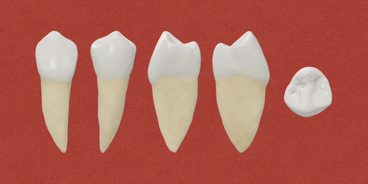
Permanent mandibular first premolar is the 4th tooth from the midline positioned posterior to the canine and anterior to first premolar. It is a transitional tooth between the anterior and posterior teeth and primarily assists them in tearing and grinding of food.
The main functions of mandibular first premolar include:
- Tearing and piercing of food.
- Initial grinding and transfer of food from canines to the molars.
- Maintaining aesthetics and vertical dimensions of face
The tooth also presents characteristics similar to both mandibular canine and mandibular first premolar.
Characteristics similar to Mandibular Canine
- Buccal aspect: Sharp and long buccal cusp. Also, it is the only functional cusp, and the mesiobuccal cusp ridge is shorter than the distobuccal cusp ridge.
- Lingual aspect: Very small lingual cusp (sometimes similar to a large cingulum). As a result, the occlusal surface slopes lingually in cervical direction.
- Proximal aspect: The buccolingual measurement is similar to that of canine.
- Occlusal aspect: The crown outline is usually diamond-shaped similar to that of canine.
Characteristics similar to Mandibular Second Premolar
- Buccal aspect: The crown and root outline resembles that of lower 2nd premolar, except for the longer buccal cusp.
- The mesial and distal contact areas are nearly at the same level.
- Proximal aspect: The curvature of cervical line on mesial and distal side are similar to that seen in lower 2nd premolar.
- The tooth presents two cusps, although the lingual cusp is non-functional.
Chronology
- First evidence of calcification: 21-24 months.
- Enamel completion: 5-6 years.
- Eruption: 10-12 years.
- Root completion: 12-13 years.
Dimensions
- Overall length: 22.5 mm
- Crown length: 8.5 mm
- Root length: 14.0 mm
- Mesiodistal diameter of crown: 7.0 mm
- Mesiodistal diameter of crown at cervix: 5.0 mm
- Buccolingual diameter of crown: 7.5 mm
- Buccolingual diameter of crown at cervix: 6.5 mm
- Curvature of cervical line: 1.0 mm (Mesial), 0.0 mm (Distal)
Surfaces, Line Angles, Point Angles
- Surfaces (5): Buccal, Lingual, Mesial, Distal, Occlusal
- Line Angles (8): Mesiobuccal, Distobuccal, Mesiolingual, Distolingual, Buccoocclusal, Distoocclusal, Linguoocclusal, Mesioocclusal
- Point Angles (4): Mesiobuccoocclusal, Distobuccoocclusal, Mesiolinguoocclusal, Distolinguoocclusal
Buccal Aspect
Crown Morphology
- Surface: The buccal surface is smooth and convex with well developed buccal cusp and prominent buccal cusp ridge. The buccal cusp ridge extends from the cusp tip to the cervical line and may present developmental depression on either side. These developmental depression separates the three buccal lobes.
- Outline/Shape: Trapezoidal with the shorter side towards the cervix. Also, the crown appears bilaterally symmetrical.
- Mesial outline: It is straight or slightly concave near the cervix, and becomes convex as it joins the mesiobuccal cusp ridge. The mesial contact area represented by the crest of curvature is just occlusal to the centre of the crown.
- Distal outline: It is also slightly concave near the cervical line, followed by convexity towards the distobuccal cusp ridge. The distal contact area is broader than the mesial contact, but both are at the same level.
- Occlusal outline: It is formed by the buccal cusp tip and its mesial and distal slopes or cusp ridges. The buccal cusp tip is sharp and pointed, and in most cases, it is located little mesial to the centre of the crown. The mesial and distal slopes of buccal cusp exhibit some concavity, unless they are worn off due to function. The mesial slope is shorter than the distal slope.
- Cervical outline: It is formed by the cervical margin and shows little curvature towards the apex. The crest of curvature usually approaches the centre of the root buccally.
Root Morphology
- The root outline resembles closely with that of mandibular canine, however, it is 3-4 mm shorter than the root of mandibular canine.
Lingual Aspect
Crown Morphology
- The crown is trapezoidal in shape with outlines similar to that seen from the buccal side.
- However, as the lingual cusp is very small, the lingual surface presents several unique features. The crown converges or tapers markedly towards the lingual side, making the most of mesial and distal surfaces visible. Also, the occlusal surface slopes lingually in cervical direction, so, most of the occlusal surface is visible.
- The lingual cusp is short and less developed, but presents a pointed cusp tip. The cusp tip is in alignment with the buccal triangular ridge.
- The cervical portion of the crown is narrow, while the contact areas and marginal ridges are pronounced and extend out above the cervical portion.
- Mesiolingual Developmental Groove: It is a characteristic feature found on mandibular first premolar. The groove extends the mesial fossa on the occlusal surface to the lingual surface and acts as a line of demarcation between the mesiobuccal lobe (mesial marginal ridge) and lingual lobe.
Root Morphology
- The root is narrower, and presents a narrow ridge (smooth and convex) along its full length. As a result, the mesial and distal surfaces of the root are visible.
- It tapers evenly from the cervix to the apex (pointed).
- Developmental depressions are often seen with developmental grooves on the mesial side.
Mesial Aspect
Crown Morphology
- Surface: The mesial surface is smooth, except for the mesiolingual groove. It is convex at the mesial contact area (located below the buccal cusp tip), and immediately below this convexity, the surface is sharply concave between the mesial contact area and the cervical line.
- Cusps: The buccal cusp tip is usually centred over the root, while the lingual cusp tip is on a line approximately with the lingual border of the root. Also, the curvature of the lingual cusp outline is lingual to the root outline. This is in contrast to the maxillary posterior teeth where both buccal and lingual cusp tips are within the confines of the root trunks.
- Outline/Shape: The crown outline is roughly rhomboidal (characteristic of all mandibular posterior teeth).
- Buccal outline: It shows marked convexity from the cervix to the buccal cusp tip and the crest of curvature is in the cervical third of the crown. This accentuated convexity and location of the crest of contour are characteristic of the buccal surface of all lower posterior teeth.
- Lingual outline: It is also curved, but shows less convexity than the buccal outline and the crest of curvature is located more occlusally in the middle third of the crown.
- Occlusal outline: It is formed by the mesiobuccal cusp ridge, mesial marginal ridge (MMR) and the mesiolingual cusp ridge. The MMR slopes lingually in the cervical direction, and merges with the developmental depression harbouring the mesiolingual developmental groove.
- Cervical outline: It is curved in occlusal direction with the crest of curvature centred buccolingually.
Root Morphology
- The root surface is smooth and flat from the buccal margin to the centre, and then converges sharply toward the root centre lingually. A deep developmental groove is often seen in this area.
- There are usually shallow grooves, however, sometimes a deep developmental groove is found ending in a bifurcation at the apical third.
- The root appears tapered from the cervix and ends in a relatively pointed apex. The apex is in line with the tip of the buccal cusp tip.
- Buccal outline of the root is curved, while the lingual outline may be straight.
Distal Aspect
Crown Morphology
- Surface: It is mostly smooth and convex, with a linear concavity just occlusal to the cervical line. Similar to the mesial side, the distal contact area is positioned below the buccal cusp tip, however, it is broader than the mesial contact area. The centre of distal contact area is at a point midway between the buccal and lingual crest of curvature and midway between the cervical line and buccal cusp tip.
- Outline/Shape: The crown is rhomboidal in shape like the mesial side, with similar buccal and lingual outlines but differing occlusal and cervical outlines.
- Buccal outline: It shows marked convexity in the cervical portion and then becomes less convex or almost straight towards the buccal cusp tip. The crest of curvature is in the cervical third.
- Lingual outline: It is less convex, but shows more uniform curvature from the cervix to the lingual cusp tip. The crest of curvature is in the middle third of the crown.
- Occlusal outline: It is formed by the distobuccal cusp ridge, distal marginal ridge (DMR) and distolingual cusp ridge. The DMR is perpendicular to the long axis of the tooth, rather than sloping lingually like the MMR. Also, the DMR is confluent with the lingual cusp and shows no developmental groove.
- Cervical outline: It is almost straight with no curvature.
Root Morphology
- The root surface shows more convexity than found medially.
- A shallow developmental groove is found at the centre of the root, but it rarely contains a deep developmental groove.
- The distal surface slopes from the buccal margin toward the centre of the root lingually, but the slope is more gradual than the mesial side.
Occlusal Aspect
-
Outline/Shape: It shows considerable variation in gross outline. Usually, it is diamond-shaped similar to the incisal aspect of mandibular canine, however, some may have a circular outline similar to the mandibular 2nd premolar.
-
Mesiobuccal and distobuccal line angles are prominent and rounded.
-
Curvature of mesial and distal contact areas are relatively broad, with the distal contact area broader of the two.
-
Crown converges sharply to the centre of the lingual surface starting from the mesial and distal contact areas. This gives a triangular appearance, with the base at the buccal cusp ridges/line angles and the apex at the lingual cusp.
-
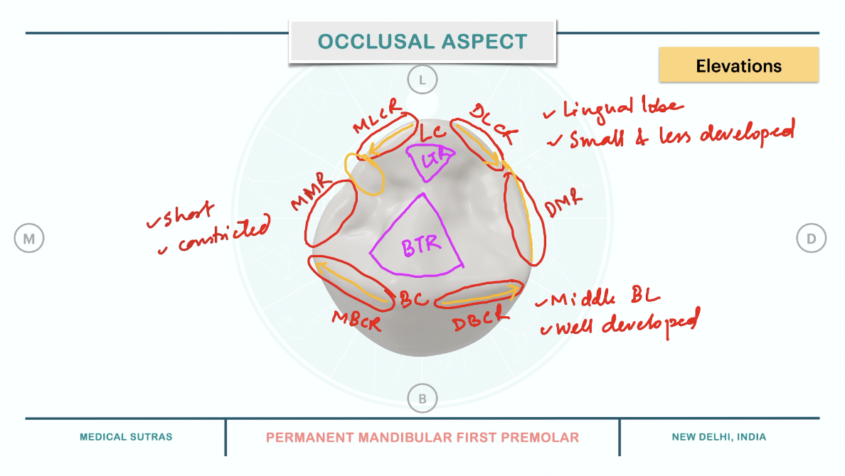
Elevations found on the occlusal surface:
- Buccal cusp: It develops from the middle buccal lobe and is well formed and makes the bulk of the crown.
- Lingual cusp: It develops from the lingual cusp, and is less developed than the buccal cusp.
- Mesiobuccal cusp ridge: Extends from the buccal cusp to the mesiobuccal line angle.
- Distobuccal cusp ridge: Extends from the buccal cusp to the distobuccal line angle.
- Mesiolingual cusp ridge: Extends from the lingual cusp to the mesiolingual line angle.
- Distolingual cusp ridge: Extends from the lingual cusp to the distolingual line angle.
- Buccal triangular ridge: It is prominent and heavy and extends from the buccal cusp to the centre of occlusal surface.
- Lingual triangular ridge: It is small and extends from the lingual cusp towards the centre of occlusal surface.
- Mesial marginal ridge: It is short and constricted, and extends from the mesiobuccal line angle to the Mesiolingual Developmental groove.
- Distal marginal ridge: It is larger than the MMR and is continuous with the distolingual cusp ridge.
-
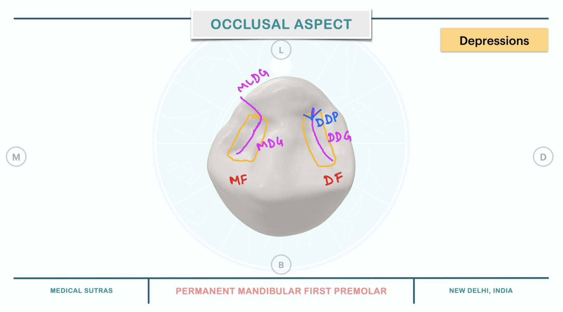
Depressions found on the occlusal surface:
- Mesial fossa: Present near the MMR. It is linear in shape and contains the mesial developmental groove.
- Distal fossa: Present mesial to DMR. It is more circular in outline and contains the distal developmental groove.
- Mesial developmental groove: It is almost straight and extends buccolingually merging with mesiolingual developmental groove.
- Distal developmental groove: It is crescent-shaped and may contain the distal developmental pit.
- Distal developmental pit: It presents several accessory supplemental grooves extending from it.
References
- Wheeler's Dental Anatomy, Physiology and Occlusion(2019), Stanley J. Nelson DDS MS, Elsevier.
*This article is excerpt from the above mentioned book and Medical Sutras does not make any ownership and affiliation claims.