Pathologic calcification involves an abnormal deposition of calcium salts, together with smaller amounts of iron, magnesium and other minerals.
It can occur in two ways:
- Dystrophic calcification &
- Metastatic calcification.
Morphology
- Gross examination: Calcium salts are seen as fine white granules or clumps, often felt as gritty deposits.
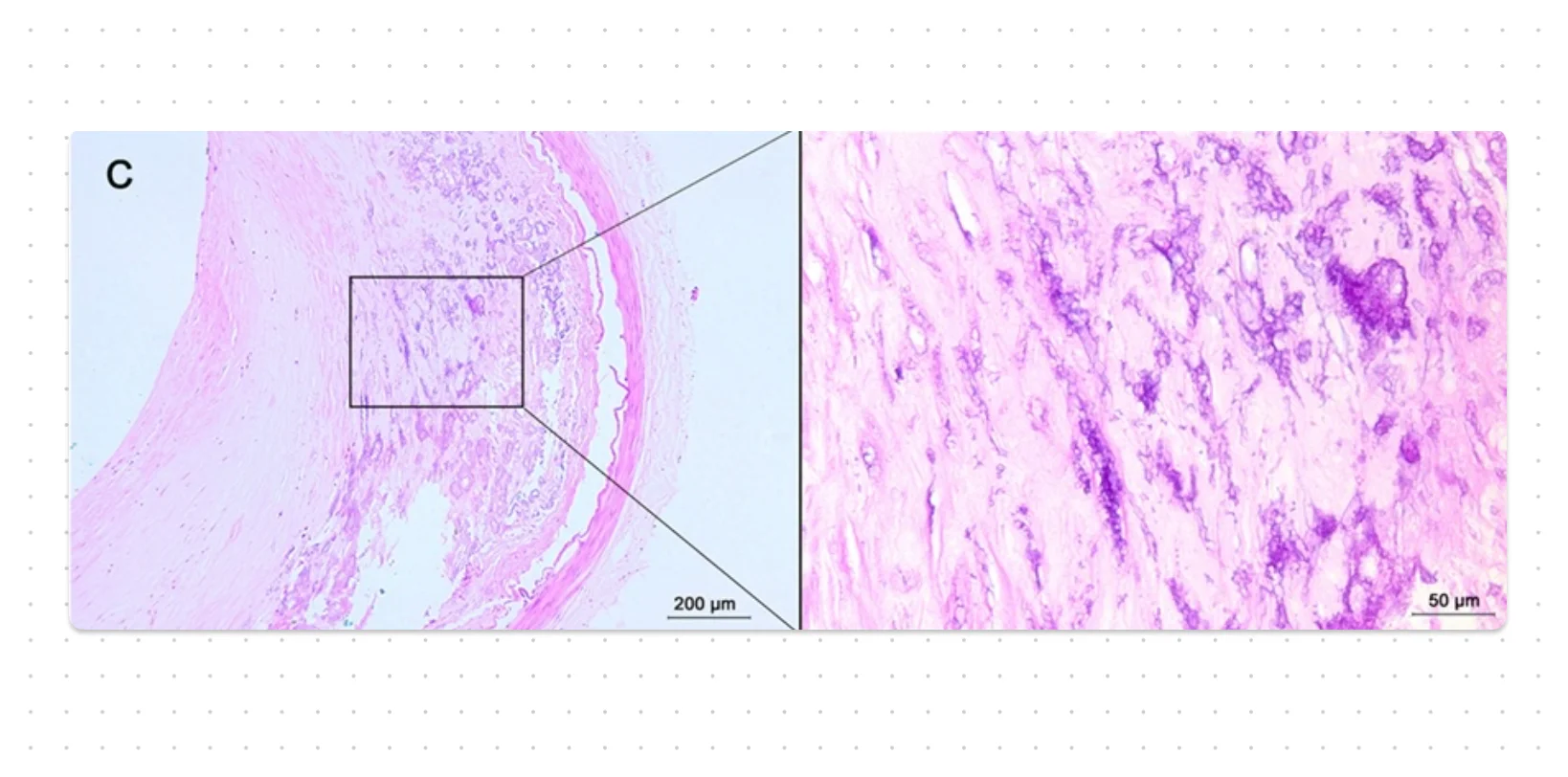
- Histologic examination: Appear as intracellular and/or extracellular basophilic deposits, and with time, there may be formation of heterotopic bone in foci of calcification.

*This file is licensed under the Creative Commons Attribution-Share Alike 4.0 International license. Author: Department of Pathology, Calicut Medical College. Source: Wikimedia Commons.
Dystrophic Calcification
There is normal calcium metabolism, but, it deposits in injured or dead tissue, such as areas of necrosis (any type).
Pathogenesis: It starts with extracellular deposition of crystalline calcium phosphate in membrane-bound vesicles. These crystals then propagate and form larger deposits.
- These may be derived from injured cells or the intracellular deposition of calcium in the mitochondria of dying cells.
- Extracellular calcium is concentrated in vesicles due to its affinity for membrane phospholipids.
- Phosphates accumulate as a result of the action of membrane-bound phosphatases.
Clinical Presentation
- It is almost universally found in the arterial lesions of advanced atherosclerosis.
- Common in areas of caseous necrosis in TB, with sometimes a tuberculous lymph node being essentially converted to radiopaque stone.
- It may be an incidental finding indicating insignificant past cell injury, or, it may be a cause of organ dysfunction, such as in aging or damaged heart valves causing severely compromised valve motion.
Metastatic Calcification
It is associated with hypercalcemia and can occur in normal tissues.
Etiology
- Increased secretion of parathyroid hormone: Due to either primary parathyroid tumors or production of parathyroid hormone-related protein by other malignant tumors.
- Destruction of bone: Due to the effects of accelerated turnover (e.g., Paget disease), immobilisation, or tumors (increased bone catabolism associated with multiple myeloma).
- Vitamin D-related disorders incl. vitamin D intoxication and sarcoidosis ( macrophages activate vitamin D precursor).
- Renal failure: Phosphate retention leads to secondary hyperparathyroidism.
Clinical Presentation
- Can occur widely throughout the body.
- Mainly affects the interstitial tissues of the vasculature, kidneys, lungs, and gastric mucosa.
- Generally do not cause clinical dysfunction. However, extensive calcifications in the lungs (radiographically evident) may produce respiratory deficits and in the kidney (nephrocalcinosis) can lead to renal damage.
References
- Robbins Basic Pathology, 10th edition, Vinay Kumar, Abul K. Abbas, Jon C. Aster, Elsevier.
- The image used in the cover photo is licensed under the Creative Commons Attribution 4.0 International license. Source: "Histological Characteristics of Intracranial Atherosclerosis in a Chinese Population: A Postmortem Study". Frontiers in Neurology 8. DOI:10.3389/fneur.2017.00488. ISSN 1664-2295.
*This article is an excerpt from the above mentioned book and Medical Sutras does not make any ownership or affiliation claims.