Osteogenesis Imperfecta, commonly manifested with fragility of bones, results from abnormality in Type I collagen i.e, failure of fetal collagen to transform into mature collagen. It involves both qualitative (abnormal collagen) and quantitative (decreased collagen) defects.
There is mutation in the loci coding for pro alpha-1 and pro alpha-2 chains of Type I collagen.
- COL1A1 on band 17q21.
- COL1A2 on band 7q22.1.
Clinical Features
- Extreme fragility and porosity of the bones.
- Increased proneness to fracture (Fracture heals rapidly but new bone is of similar imperfect quality).
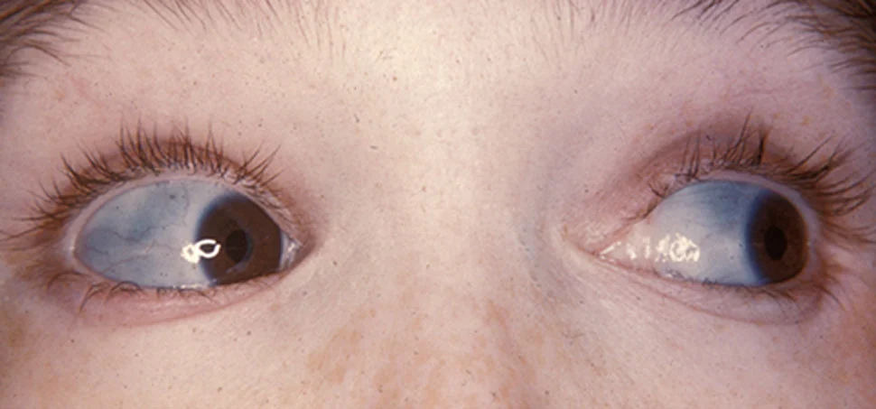
- Pale, blue sclerae (Abnormally thin sclerae - pigmented choroid show through, producing the bluish color).
- Deafness due to osteosclerosis.
- Abnormalities of teeth (Hereditary opalescent dentin).
- Laxity of ligaments.
- Peculiar shape of the skull.
- Abnormal electrical reaction of the muscle.
- Tendency towards capillary bleeding.
- Congenital or Vrolik's type : Present at birth.
- Tarda or Lobstein's type : Recognized later in life, also called osteopsathyrosis.
Type I Osteogenesis Imperfecta
- Most common and mildest form.
- Blue sclera.
- In-utero fractures in 10% of cases.
- Mild-to-moderate bone fragility, decreases after puberty.
- Kyphoscoliosis.
- Hearing loss.
- Short stature.
- Subtype A : Dentinogenesis Imperfecta is absent.
- Subtype B : Dentinogenesis Imperfecta is present.
Type II Osteogenesis Imperfecta
- Exhibits extreme fragility ad frequent fractures.
- In-utero fracture in 100% of cases.
- Most are still born.
- 90% die before 4 weeks of age.
Type III Osteogenesis Imperfecta
- In-utero fracture in 50%, remaining 50% in neonatal period.
- Associated with Dentinogenesis Imperfecta.
- Sclera is of variable hue.
- Limb shortening and progressive deformities.
- Triangular facies and frontal bossing.
- Pulmonary hypertension.
- Occasional hearing loss.
Type IV Osteogenesis Imperfecta
- Normal sclera and occasional hearing loss.
- Fractures begin in infancy.
- Mild angulation and shortening of long bones.
- Subtype A : Dentinogenesis Imperfecta is absent.
- Subtype B : Dentinogenesis Imperfecta is present.
Oral Findings
- Hypoplasia of dentin and pulp, translucent teeth with yellow or blue-gray coloration, delayed tooth eruption and susceptibility to caries.
- Class III malocclusion and maxillary hypoplasia.
- Large number of impactions and ectopic teeth.
- Exaggerated occiput.
- Frontal and temporal bossing.
- Large head size.
Radiographic Findings
- Osteopenia.
- Bowing, Angulation or deformity of long bones.
- Wormian bones (Sutural bones).
Points to Note
-
Also known as Brittle bone, Lobstein's disease.
-
It represents a hereditary autosomal trait.
-
Osteochondrodysplasia : Abnormalities of cartilage or bone growth and development (generalized disorder).
-
Dysostoses : Malformations of individual bones, single or in combination.
-
Blue Sclera also found in :
- Osteoporosis
- Fetal Rickets
- Turner Syndrome
- Paget's Disease
- Marfan Syndrome
- Ehlers' Danos Syndrome
- Normal Infants.
References
-
Shafer, Hine, Levy Shafer's Textbook of Oral Pathology (7th Edition), Editors - R Rajendran, B Sivapathasundharam, Elsevier.
-
Textbook of Oral Medicine (3rd Edition), Anil Govindrao Ghom, Savita Anil Ghom (Lodam), Jaypee Brothers Medical Publishers (P) Ltd.
-
Martin S. Greenberg, Michael Glick, Jonathan A. Ship - Burket's Oral Medicine, 11th Edition (2008), BC Decker Inc.
-
The image used is licensed under the Creative Commons Attribution License (CC-BY 2.0).
- Description : The classic blue sclera of a person with osteogenesis imperfecta.
- Source : http://cnx.org/contents/_on798ZB@3/Images-of-Memorable-Cases-Case
- Author : Herbert L. Fred, MD and Hendrik A. van Dijk.
*This article is an excerpt from the above mentioned books and Medical Sutras does not make any ownership or affiliation claims.