The optic nerve is the 2nd cranial nerve that is responsible for vision.
Functional Components
- Special somatic afferent fibres for vision.
- Afferents for pupillary light and accommodation reflexes.
- Few efferent fibres, sources of origin and their functions remain unsettled.
Characteristics
- Purely sensory: Consist of special somatic afferent fibres, that carry sense of sight from the visual field of the corresponding eye.
- Not a true peripheral (cranial nerve): Actually a tract of brain that develops as an outgrowth of diencephalon during fetal life.
- Consists of second-order sensory neurons.
- Fibres are myelinated by oligodendrocytes.
- Surrounded by meninges.
- Fibres cannot regenerate if cut/damaged.
Anatomy
The optic nerve is 4 cm in length and is divided into three parts:
- Intraorbital part.
- Canalicular part.
- Intracranial part.
It is enclosed by three meninges of the brain:
- The thick fibrous dural sheath of optic nerve blends with the sclera of the eyeball.
- The subarachnoid space containing CSF surrounds the optic nerve and is continuous with the subarachnoid space of the brain.
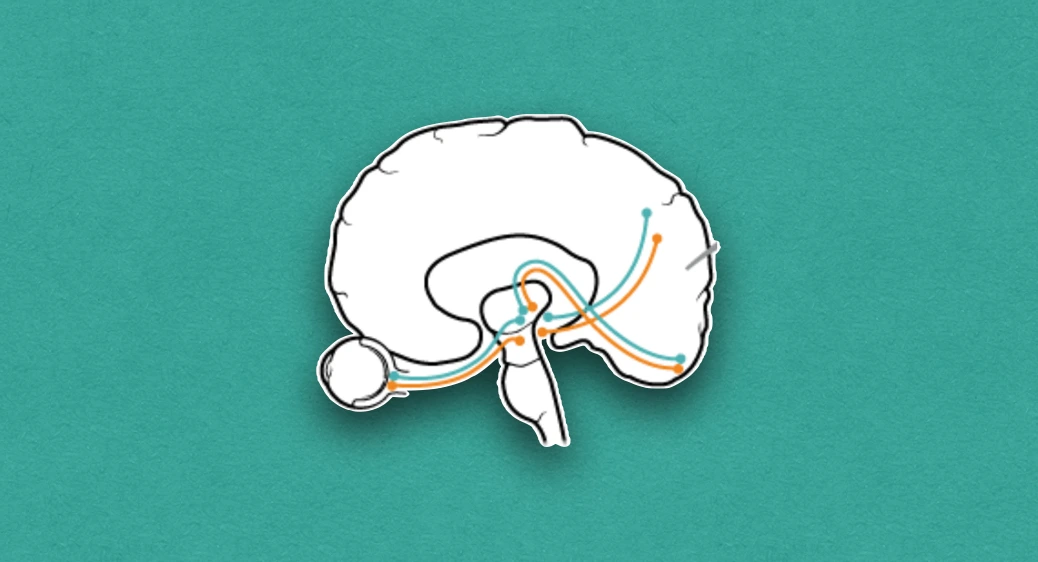
Pathway
- The optic nerve fibres arise from ganglion cells in the neural layer of the retina of the eyeball.
- They converge toward the optic disc at the posterior pole of the eyeball, pierce the outer layer of retina, choroid and sclera to leave the eyeball.
- Optic nerve: Formed by the union of the fibres immediately after they emerge from the eyeball. It passes posteromedially through the posterior half of the orbit and enters the middle cranial fossa through the optic canal.
- Optic chiasma: In the middle cranial fossa, the optic nerves of both sides unite to form the optic chiasma. The mid-region of optic chiasma is composed of crossed fibres from the medial/nasal halves of the retina of both eyes, while the lateral region is made up of fibres from the lateral/temporal half of the retina of the ipsilateral eye.
- Optic tracts: Diverge from the optic chiasma. Most of the fibres of the optic tract relay in the lateral geniculate body.
- Optic radiations: The third-order neurons arise in the lateral geniculate body, run in the retrolenticular part of the internal capsule and form optic radiations. The fibres of optic radiation terminate in and around the calcarine sulcus of the occipital lobe (visual cortex).
- Some of the fibres from the lateral geniculate body reach the pretectal area of the midbrain and form a part of the pathway for light reflex.

Clinical Significance
- Ipsilateral total blindness: May occur due to damage of optic nerve or blockage of central artery of retina. The compression of optic nerve results in optic atrophy, which subsequently leads to ipsilateral total blindness called anopia.
- Papilledema: An increased CNS pressure within the cranial cavity impedes the return of venous blood from the retina by pressing the central vein of retina and its tributaries, This results in swelling of optic disc owing to edema called papilledema. It is a valuable clinical evidence of an increased intracranial pressure.
- Clinical testing of optic nerve: By performing tests for visual acuity, color perception, and loss of vision in different visual fields.
References
-
[](https://amzn.to/3Ixnict) Textbook of Anatomy Head, Neck, and Brain (Volume III), Vishram Singh
-
The image used is licensed under the Creative Commons Attribution 4.0 International license. (Author: OpenStax. Source: https://cnx.org/contents/FPtK1zmh@8.25:fEI3C8Ot@10/Preface)
*This article is an excerpt from the above mentioned sources and Medical Sutras does not make any ownership or affiliation claims.