Odontomas (Odontomes) are developmental malformations (hamartomas) of dental tissues, in which epithelial and mesenchymal cells exhibit complete differentiation into ameloblasts and odontoblasts, resulting in formation of enamel and dentin at abnormal places and in abnormal pattern (since the odontogenic cells fails to reach normal state of morphodifferentiation).
It is composed of more than one type of tissue, hence, also called composite odontoma.
The etiology is unknown. However, factors such as local trauma, infection and genetic mutations have been implicated in the pathogenesis of odontomas.
Classification
- Compound composite odontoma : Consists of enamel and dentin laid down in a structured manner that have considerable anatomic resemblance to well formed small teeth.
- Complex composite odontoma : Consists of non-discrete, irregular masses of calcified dental tissues that have no morphologic similarity even to rudimentary teeth.
Clinical Findings
-
Most odontomas are asymptomatic and are discovered as an accidental finding during routine radiographs.
-
Occasional findings : Unerupted or impacted teeth, retained deciduous teeth, swelling and evidence of infection.
-
Age : Adolescents and young adults.
-
Gender : Slight predilection for males.
-
Location : More common in maxilla than mandible.
- Compound odontoma : Anterior maxilla.
- Complex odontoma : Posterior mandible.
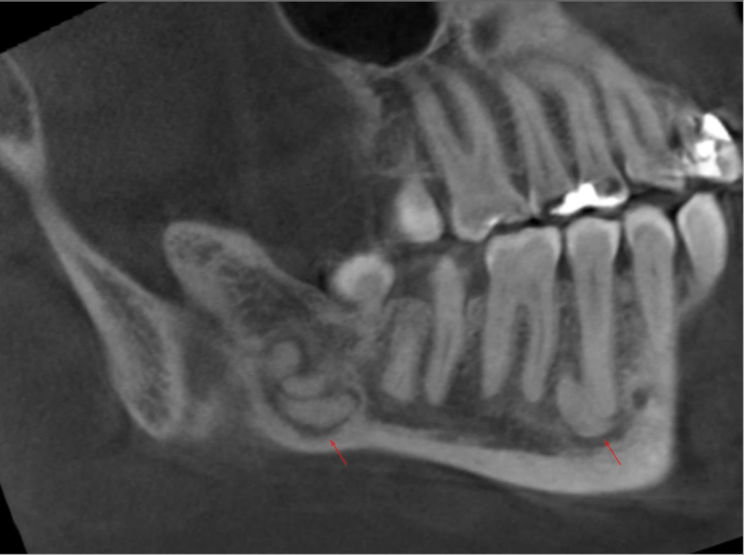
Radiographic Features
- Often seen as radiopaque mass, situated between the roots of teeth or associated with unerupted teeth.
- Compound odontomas : Cluster of multiple, small tooth-like structures surrounded by a radiolucent line (representing pericoronal space of the unerupted teeth).
- Complex odontomas : Irregular mass of calcified material surrounded by a narrow radiolucent band with a smooth outer periphery.
- The radiographic appearance may need to be differentiated from radiopaque lesions such as osteoblastoma, osteoid osteoma, enostosis and idiopathic osteosclerosis.
Histologic Features
- Normal-appearing enamel or enamel matrix, dentin, pulp tissue and cementum, that may or may not be in normal relation to one another.
- Connective tissue capsule around the odontoma is similar to the follicle surrounding a normal teeth.
- Ghost cells are also found in some cases.
Treatment
The recommended treatment is surgical removal of the odontoma and there is no recurrence after removal.
Points to Note
- Odontoma is considered the most common odontogenic tumor.
- Multiple odontomas are found in Gardner's Syndrome.
- It is suggested that all odontomas must be sent for histopathologic examination, since they bear great resemblance to ameloblastic odontoma and ameloblastic fibro-odontoma.
- Ameloblastic odontoma (Odontoameloblastoma) : It is an odontogenic neoplasm characterised by the simultaneous occurrence of ameloblastoma and odontoma in the same tumor mass.
- Ameloblastic fibro-odontoma : It is a mixed lesion consisting of odontoma and soft tissue resembling ameloblastic fibroma. If only dentin is present, it is referred as ameloblastic fibro-dentinoma.
- If odontomas are left untreated, they may give rise to cysts of dentigerous type (formed by separation of reduced enamel epithelium from enamel).
References
-
Shafer, Hine, Levy Shafer's Textbook of Oral Pathology (7th Edition), Editors - R Rajendran, B Sivapathasundharam, Elsevier.
-
Cawson's Essentials of Oral Pathology and Oral Medicine, 9th Edition, EW Odell, Elsevier.
-
Textbook of Oral Pathology, 2nd Edition, Anil Govindrao Ghom, Shubhangi Mhaske (Jedhe) - Jaypee Brothers Medical Publishers (P) Ltd.
-
Burket's Oral Medicine, 13th Edition, Michael Glick, Martin S. Greenberg, ,Peter B. Lockhart, Stephen J. Challacombe, Wiley Blackwell.
-
The image used is licensed under the Creative Commons Attribution-Share Alike 3.0 Unported license.
- Deutsch: Odontom(e) im Unterkiefer bei einer Mittvierzigerin: Dichte, amorphe Formation in der Nähe aber nicht ausgehend von den Zahnwurzeln. Befund nicht histologisch bestätigt, aber gut passend. DVT in verschiedenen Reformatierungen.
- Author : Hellerhoff.
*This article is an excerpt from the above mentioned book and Medical Sutras does not make any ownership or affiliation claims.