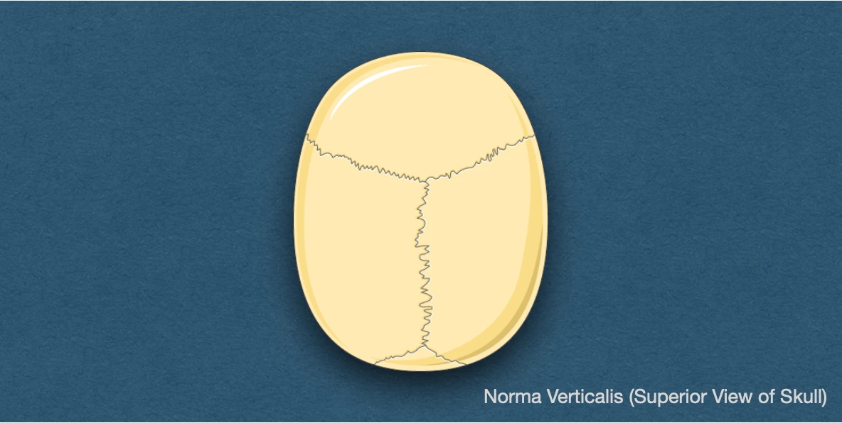
It refers to the superior view of the skull. When viewed from above, it is usually oval in shape, being wider posteriorly than anteriorly.
Bones
- Upper/Squamous part of frontal bone (anteriorly).
- Parietal bone (on each side of midline).
- Upper/Squamous part of occipital bone (posteriorly).
Sutures
- Coronal suture: It crosses the cranial vault from side-to-side and runs downwards and forwards between the posterior border of frontal bone and the anterior border of the two parietal bones.
- Sagittal suture: Lies in the median plane between the two parietal bones.
- Lambdoid suture: Shaped like the letter lambda, it runs downwards and forwards between the posterior border of the two parietal bones and the occipital bone.
- Metopic suture: Occasionally present in 3-8% cases, it separates the two halves of the frontal bone along the median plane. It represents the remnants of suture between the two frontal bones, and normally fuses at 6 years of age.
Important Features
- Vault of skull: Arched roof for the dome of skull.
- Vertex: Highest point on the skull. It lies on the sagittal suture near its middle, a few centimetres behind the bregma.
- Parietal tuber/eminence: Area of maximum convexity of the parietal bone
- Bregma: Meeting point of coronal and sagittal sutures. In the foetal skull, it presents as the anterior fontanelle, that closes at 18-24 months of age.
- Lambda: Meeting point of the sagittal and lambdoid sutures. In the foetal skull, it is the site of posterior fontanelle (closes at 2-3 months of age).
- Parietal foramen: Present on the parietal bone near the sagittal suture, 2.5-4 cm in front of lambda. It transmits emissary vein from the veins of scalp to superior sagittal sinus.
- Obelion: Point on sagittal suture between the two parietal foramina.
- Temporal lines: Present over the parietal bone, these start as a single line from the zygomatic process of frontal bone. Moving posteriorly, there are two lines that arches backward and upward, and cross the frontal bone, coronal suture and the parietal bone. The superior line fades out over the posterior part of the parietal bone, while, the inferior line continues downwards and forwards with the zygomatic arch.
References
- BD Chaurasia's Human Anatomy Volume 3 (Head & Neck), Eighth edition, CBS Publishers and Distributors Pvt Ltd
- Textbook of Anatomy Head, Neck, and Brain (Volume III), Vishram Singh
- Image Source: Smart Servier website: Rheumatology. This file is licensed under the Creative Commons Attribution-Share Alike 3.0 Unported license.
*This article is an excerpt from the above mentioned sources and Medical Sutras does not make any ownership or affiliation claims.