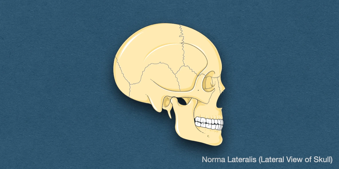
Norma lateralis refers to the lateral view of the skull, that is, from the sides.
Bones
- Frontal bone.
- Parietal bone.
- Occipital bone.
- Temporal bone
- Zygomatic bone.
- Nasal bone.
- Lacrimal bone.
- Maxilla.
- Mandible.
Important Features
Temporal Lines
- Present over the parietal bone, these start as a single line from the zygomatic process of frontal bone. Moving posteriorly, there are two lines that arches backward and upward, and cross the frontal bone, coronal suture and the parietal bone.
- Superior temporal line fades out over the posterior part of the parietal bone.
- Inferior temporal line continues downwards and forwards with the zygomatic arch (with supramastoid crest on the squamous temporal bone). It continues anteriorly with the posterior root of the zygomatic arch.
Zygomatic Arch (Zygoma)
-
It is curved, horizontal bony bar present in front of the ear, formed by the temporal process of zygomatic bone (anterior one-third) and the zygomatic process of temporal bone (posterior two-thirds).
-
The posterior end of the arch is attached to the squamous temporal bone by anterior and posterior roots.
- Anterior root passes medially in front of the articular fossa.
- Posterior root passes backwards along the lateral margin of the articular fossa, then, above the external acoustic meatus, and become continuous with the supramastoid crest.
-
Jugal point: It is a point at the anterior end of the upper border of the zygomatic arch.
-
Articular tubercle of the root of zygoma (Tuberculum articulare): It is a rounded prominence/projection found along the inferior aspect of the zygomatic process of temporal bone, at the junction of anterior and posterior roots of zygoma.
-
Postglenoid tubercle: It is a projection found along the inferior aspect of the squamous part of temporal bone, located anterosuperior to the external acoustic meatus. It forms the posterior aspect of the mandibular/articular fossa.
External Acoustic Meatus (External auditory meatus/Ear canal)
- It is a sigmoid or S-shaped tube that extends from the deep part of the concha to the tympanic membrane, and opens just below the posterior part of posterior root of zygoma.
- Boundaries: Tympanic plate (anteroinferior) and squamous temporal bone (postero-superior).
- Suprameatal triangle (Triangle of McEwen): Small, swallow depression on the squamous part of temporal bone. It is present postero-superior to the meatus and forms the lateral wall of the tympanic or mastoid antrum.
Styloid Process
- It a thin, needle-like bony projection from inferior part of the temporal bone, situated anteromedial to the mastoid process (in front of the stylomastoid foramen).
- It is directed downwards, forwards, and slightly medially.
- It provides attachment to to the stylopharyngeus, stylohyoid, and styloglossus muscles, as well as the stylohyoid and stylomandibular ligaments.
Pterion
-
H-shaped anatomic landmark on the lateral side of skull that represents the junction of four bones: Frontal, Parietal, Squamous part of temporal, and Greater wing of sphenoid bone.
-
It is located approximately 4 cm superior to the zygomatic arch and 3.5 cm posterior to the frontozygomatic suture.
-
It signifies the site of closed anterolateral/sphenoidal fontanelle.
-
According to Murphy (1956), pterion can be categorised into four types:
- Sphenoparietal (most common): Formed by the articulation of the greater wing of sphenoid bone with parietal bone
- Frontotemporal suture: Formed between the frontal and temporal bone.
- Stellate suture: Formed by the fusion of sphenoid, frontal, parietal and temporal bones
- Epipteric (wormian) suture: Characterised by the presence of small sutural bones between the sphenoid and parietal bones.
-
It overlies various anatomical structures, namely, the anterior division of the middle meningeal vessels, middle cerebral vessels, Sylvain fissure, circle of Willis, insula and Broca's motor speech area (on the left) and optic nerve
-
Any trauma in this region may injure the middle meningeal vessels, leading to extradural haemorrhage/epidural hematoma.
Asterion
- Anatomic landmark on the lateral side of the skull formed at the junction of the occipital, temporal, and parietal bone.
- It is located at the junction of the lambdoid, occipitomastoid and parietomastoid suture.
- Internally, it is related to the transverse and sigmoid sinuses.
- It signifies the site of closed mastoid fontanelle.
- It is used as a surface landmark for the radiological and anthropological measurement of the skull.
Fossae
- Mandibular/Articular fossa/Glenoid fossa.
- Temporal fossa.
- Infratemporal fossa.
- Pterygopalatine fossa.
References
- BD Chaurasia's Human Anatomy Volume 3 (Head & Neck), Eighth edition, CBS Publishers and Distributors Pvt Ltd.
- Muche A. Positions and Types of Pterion in Adult Human Skulls: A Preliminary Study. Ethiop J Health Sci. 2021 Jul;31(4):875-884. doi: 10.4314/ejhs.v31i4.23. PMID: 34703188; PMCID: PMC8512946. https://pmc.ncbi.nlm.nih.gov/articles/PMC8512946/
- Cowan PT, Sandhu G, Adigun OO. Anatomy, Head and Neck: Asterion. [Updated 2023 Feb 20]. In: StatPearls [Internet]. Treasure Island (FL): StatPearls Publishing; 2024 Jan-. Available from: https://www.ncbi.nlm.nih.gov/books/NBK537214/
- Textbook of Anatomy Head, Neck, and Brain (Volume III), Vishram Singh
- Image Source: Smart Servier website: Rheumatology. This file is licensed under the Creative Commons Attribution-Share Alike 3.0 Unported license.
*This article is an excerpt from the above mentioned sources and Medical Sutras does not make any ownership or affiliation claims.