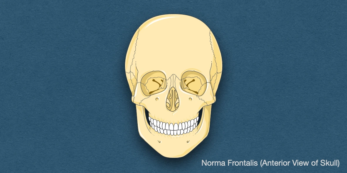
Norma frontalis refers to the anterior/frontal view of the skull, that is roughly oval in shape, being wider above than below.
It can be divided into the following regions:
- Frontal region
- Orbital openings
- Anterior nasal aperture
- Zygomatic bones (Malar bones)
- Maxilla/Upper jaw
- Lower jaw/mandible
Frontal Region
It is formed by the squamous part of the frontal bone and presents:
- Superciliary arch: Rounded, curved elevation just above the medial part of each orbit, overlying the frontal sinus. It is better marked in males than in females.
- Glabella: Median elevation that connects the two superciliary arches.
- Nasion: Median point at the root of the nose where the internasal suture meets with frontonasal suture.
- Frontal tuber/eminence: Low rounded elevation above the superciliary arch, on each side. It is more prominent in females and children.
Orbital Openings
These refers to the openings of the orbital cavities, present above and lateral to the anterior nasal aperture.
Boundaries
The orbital openings are quadrangular in shape and presents the following boundaries:
- Supraorbital margin: Formed entirely by the frontal bone, it presents the supraorbital notch or foramen, at the junction of medial one-third and lateral two-third. The foramen transmits the supraorbital nerves and vessels.
- Infraorbital margin: Formed by the zygomatic bone (laterally) and maxilla (medially).
- Medial orbital margin: Formed by the frontal bone above, and the lacrimal crest of the frontal process of maxilla below.
- Lateral orbital margin: Formed by the frontal process of zygomatic bone (inferiorly), and zygomatic process of frontal bone (superiorly). The bones meet at the frontozygomatic suture.
Anterior Nasal Aperture (Piriform aperture)
It is pear-shaped, being wide below and narrow above.
Boundaries
- Superior: Lower border of nasal bones.
- Inferior and Lateral: Nasal notch of the maxillae (on each side).
Features
- Anterior nasal spine: Sharp bony projection present in the median plane (lower bober of the piriform aperture) formed by the fusion of the two maxillary bones at the intermaxillary suture.
- Rhinion: Lowermost point of the internasal suture.
Articulation of the Nasal Bones
- Anteriorly: Opposite nasal bone (Internasal suture).
- Posteriorly: Frontal process of maxilla.
- Superiorly: Frontal bone (Frontonasal suture).
- Inferiorly: Upper nasal cartilage.
Maxilla (Upper jaw)
The maxillary bones presents the following features in the frontal view:
-
Nasal notch: Deep indentation along the anteromedial border of the maxilla, that forms the lateral concave margin/rim of the anterior nasal aperture.
-
Anterior nasal spine: Sharp bony projection present in the median plane (lower bober of the piriform aperture) formed by the fusion of the two maxillary bones at the intermaxillary suture.
-
Infraorbital foramen: Present 1 cm below the infraorbital margin. It transmits infraorbital nerves and vessels.
-
Incisive fossa: Above the incisor teeth.
-
Canine fossa: Lateral to the canine eminence.
-
Three processes of the maxillary bone
- Frontal process: Directed upwards, and articulates anteriorly with the nasal bone, posteriorly with the lacrimal bone, and superiorly with the frontal bone.
- Zygomatic process: Short and stout (bulky), articulates with the zygomatic bone.
- Alveolar process: Presents sockets for teeth of upper jaw.
Zygomatic Bones
Situated on the lower and lateral side of orbit, they form the prominence of the cheek.
Features
- Zygomaticofacial foramen: It is a small opening on the lateral/facial surface near the orbital border. It transmits the zygomaticofacial branch of maxillary nerve.
Mandible (Lower jaw)
- Alveolar arch: Formed by the upper border of the alveolar process, it presents the sockets for lower teeth.
- Gonion: It is a point on the posteroinferior corner/angle of the mandible.
- Mental protuberance: It is the triangular, midline elevation found along the external surface of the body of the mandible, that contributes to the formation of the prominence of chin. It continues superiorly with the mandibular symphysis, and laterally, with the mental tubercles.
- Symphysis menti (Mental symphysis or mandibular symphysis): It is a vertical, sometimes indistinct, line found along the midline on the body of the mandible, that marks the site of fusion between the right and left halves of the mandible.
- Mental point or Gnathion: It is the most antero-inferior point on the symphysis (mid-point along the base of the mandible).
- Mental tubercle (Genial tubercle): Refers to the prominence on each side of the mental protuberance, near the lower border of mandible.
- Mental foramen: Located on the external surface of the mandible, between the roots of the first and second premolars, or below the second premolar. It transmits the mental nerve and vessels.
- Oblique line (External oblique ridge): Bony ridge of the external surface, that runs upwards and backwards from the mental tubercle to the anterior border of the ramus of the mandible. It provides an origin site for the depressor anguli oris muscle.
Bones
- Frontal bone.
- Maxillae.
- Nasal bones.
- Zygomatic bones.
- Mandible.
Sutures
- Frontonasal suture.
- Frontomaxillary suture.
- Zygomaticofrontal suture.
- Zygomaticomaxillary suture.
- Lacrimomaxillary suture: It is a cranial suture between the maxilla and the anterior margin of the lacrimal bone.
- Nasomaxillary suture.
- Internasal suture.
- Intermaxillary suture.
References
- BD Chaurasia's Human Anatomy Volume 3 (Head & Neck), Eighth edition, CBS Publishers and Distributors Pvt Ltd
- Textbook of Anatomy Head, Neck, and Brain (Volume III), Vishram Singh
- Image Source: Smart Servier website: Rheumatology. This file is licensed under the Creative Commons Attribution-Share Alike 3.0 Unported license.
*This article is an excerpt from the above mentioned sources and Medical Sutras does not make any ownership or affiliation claims.