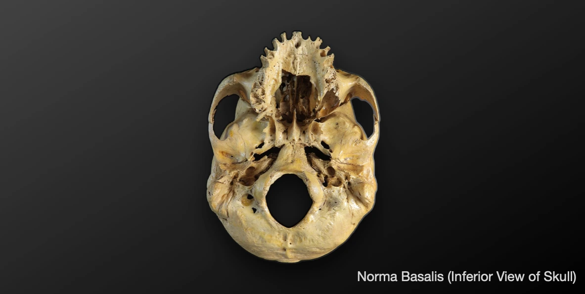
Norma basalis refers to the inferior view of skull. It can be divided into anterior, middle and posterior parts by two imaginary lines.
- Anterior transverse line: Passes along the posterior free margin of hard palate.
- Posterior transverse line: Passes along the anterior margin of foramen magnum.
Anterior Part
It lies anterior to the posterior free margin of hard palate, and consists of the alveolar processes of maxilla and hard palate.
Bones
- Maxillae.
- Palatine bones.
Sutures
- Intermaxillary suture.
- Interpalatine suture.
- Palatomaxillary suture.
Features
-
Alveolar arch: It a U-shaped ridge of bone formed by the alveolar processes of maxillae.
-
Hard palate: It is formed by the palatine processes of maxillae (anterior two-third) and horizontal plates of palatine bones (posterior one-third).
- Incisive foramen: Deep fossa situated anteriorly in the median plane. It is pierced by two incisive canals (right and left).
- Greater palatine foramen: Present just behind the lateral part of the palatomaxillary suture. These are one on each side, and provides passage to anterior palatine nerve.
- Lesser palatine foramen: 2 or 3 in number, located behind the greater palatine foramen. They perforate the pyramidal process of palatine bone, and provide passage to middle and posterior palatine nerves.
- Posterior nasal spine: Present in the midline along the posterior border of hard palate.
- Palatine crest: Curved ridge near the posterior border of hard palate, that runs medially from behind the greater palatine foramen.
Middle Part
It is the area between the posterior border of hard palate and the imaginary transverse line passing through anterior margin of the foramen magnum.
Bones
- Vomer: It is a single, thin, quadrilateral bone, located along the midline between the right and left nasal cavities. The median area of the middle part of norma basalis presents the posterior border of vomer (separates the two posterior nasal apertures).
- Sphenoid bone: Infratemporal surface of greater wing of sphenoid, pterygoid processes and spine of sphenoid.
- Occipital bone: The basilar part of occipital bone forms a broad bar of bone by fusion with the body of sphenoid.
- Temporal bone (petrous part).
Features
-
Choanae (Posterior/Internal nasal aperture): The two posterior nasal apertures are separated by the posterior border of vomer.
-
Pharyngeal tubercle: It is present on the broad bar of bone formed by sphenoid and occipital bones.
-
Pterygoid process of sphenoid bone: Projects downward from the junction of greater wing and body of sphenoid bone, behind the third molar. Inferiorly, it divides into two plates that are fused anteriorly, while divided posteriorly by the pterygoid fossa.
- Medial pterygoid plate: It is directed backwards and has a free posterior border. The upper end of posterior border divides to enclose a triangular depression called scaphoid fossa, while the lower end is prolonged downwards and laterally to form ptreygoid hamulus.
- Lateral pterygoid plate: It is directed backwards and laterally. Sometimes, the posterior border shows a projection at the middle, called pterygospinous process.
-
Spine of sphenoid: Small, sharp bony projection posterolateral to the foramen spinousum.
-
Sulcus tubae (Groove for auditory tube): It is the groove between the posteromedial margin of greater wing of sphenoid and petrous temporal bone. It lodges the cartilaginous part of auditory tube.
Foramens
- Foramen Ovale: Large, oval in shape, situated posterolateral to the upper end of posterior border of lateral pterygoid plate.
- Foramen Spinosum: Small, circular, situated posterolateral to foramen ovale.
- Foramen of Vesalius (Emissary sphenoidal foramen): Located. between foramen ovale and scaphoid fossa
- Canaliculus innominatus: Very small foramen present between foramen ovale and spinosum.
- Carotid foramen: External opening of the carotid canal, present on the inferior surface of petrous temporal bone. It transmits the internal carotid artery.
- Foramen Lacerum: Short, wide canal 2 cm long, located in the petrous temporal bone.
Posterior Part
It is the part posterior to the anterior margin of foramen magnum.
Bones
- Occipital bone.
- Temporal bone.
Sutures
- Occipitotemporal suture.
Features (Median Area)
- Foramen magnum: Largest foramen of skull, it opens upward into the posterior cranial fossa, and downward into the vertebral canal.
- External occipital crest: Begins at the posterior margin of foramen magnum, and ends posteriorly and above at the external occipital protuberance.
- External occipital protuberance: Bony projection located at posterior end of crest.
- Nuchal lines: It includes the superior and inferior nuchal lines, and occasionally, highest nuchal lines.
Features (Lateral Area)
- Occipital condyles: Oval shaped processes, one on each side of anterior part of foramen magnum. They articulate with the superior articular facets of the atlas vertebra (atlanto-occipital joints).
- Hypoglossal/Anterior condylar canal: Located anterosuperior to occipital condyle.
- Posterior condylar canal: Present occasionally in the floor of condylar fossa behind the occipital condyle.
- Condylar fossa: Small fossa located behind the occipital condyle. Sometimes, it is perforated by condylar (posterior condylar) canal.
- Jugular process of occipital bone: Lies lateral to occipital condyle and forms the posterior boundary of jugular foramen.
- Jugular foramen: Large elongated foramen located at the posterior end of petro-occipital suture.
- Jugular fossa: Deep depression on the underside of petrous temporal bone, at the posterior end of jugular foramen. It lodges the superior bulb of the internal jugular vein.
- Tympanic canaliculus: Opens on or near the thin edge of bone between the jugular fossa and lower end of carotid canal.
- Stylomastoid foramen: Located between the roots of styloid and mastoid processes, it transmits facial nerve and stylomastoid artery.
References
- BD Chaurasia's Human Anatomy Volume 3 (Head & Neck), Eighth edition, CBS Publishers and Distributors Pvt Ltd
- Textbook of Anatomy Head, Neck, and Brain (Volume III), Vishram Singh
- Image Source: Wikimedia Commons. Author: MAKY.OREL. This file is made available under the Creative Commons CC0 1.0 Universal Public Domain Dedication.
*This article is an excerpt from the above mentioned sources and Medical Sutras does not make any ownership or affiliation claims.