The human body is constructed from four fundamental tissue types—epithelial, connective, muscular, and nervous tissues. These primary tissues structurally and functionally integrate to form complex, specialized organs, relying on precise cellular interactions, specialized interfaces like the basal lamina, and diverse extracellular matrices to maintain physiologic homeostasis and enable complex activities.
🔍 Detailed Breakdown
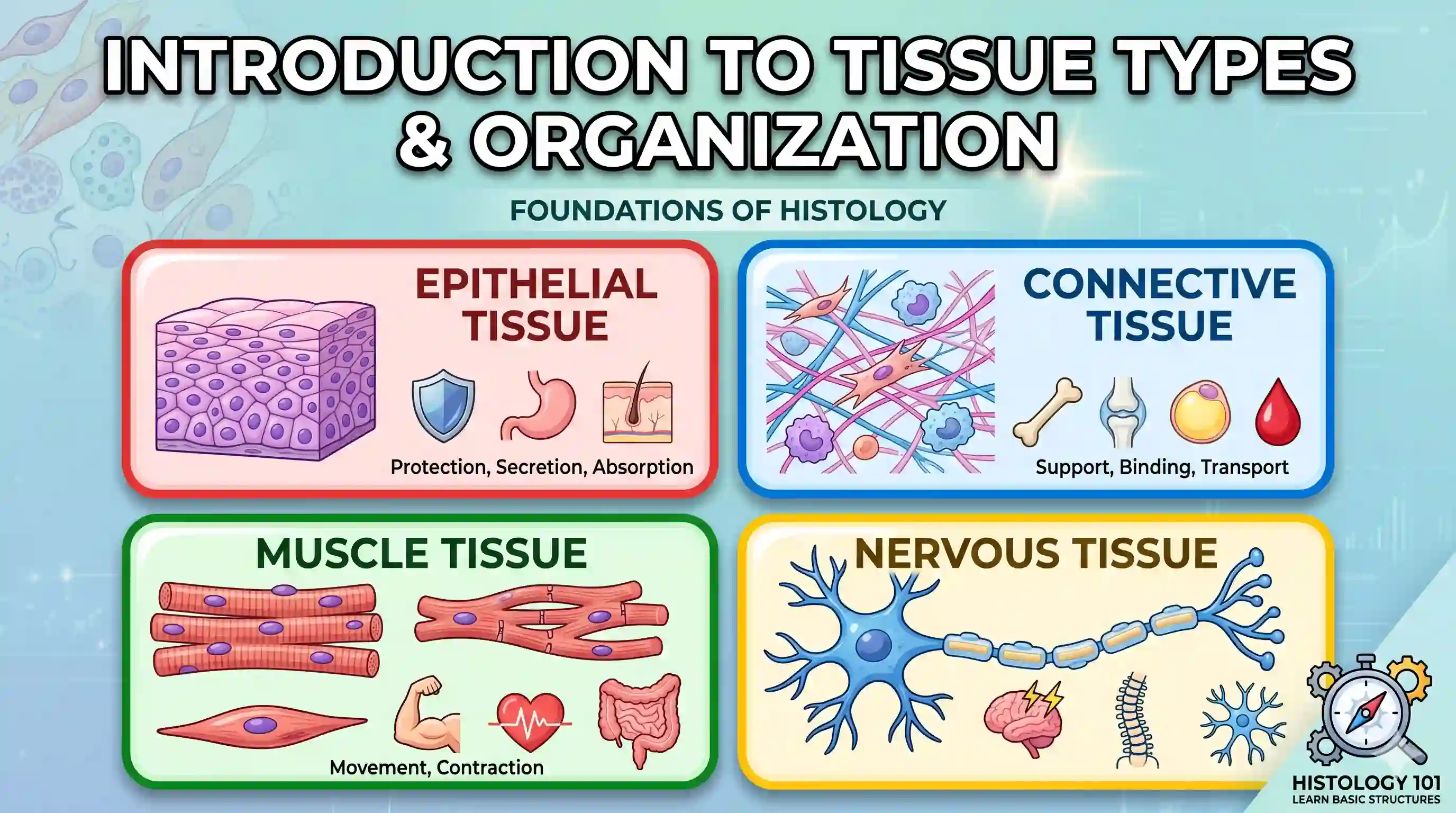
- Epithelial Tissue: Functions primarily as a highly cohesive protective barrier (e.g., stratified squamous oral epithelium) or as active secretory units (e.g., salivary gland acini). Epithelial cells (such as keratinocytes) are tightly bound by intercellular junctions (desmosomes, tight junctions, gap junctions) and are entirely avascular, relying on diffusion from adjacent tissues for nutrition.
- Connective Tissue: Provides structural scaffolding, metabolic support, and immune defense. It consists of cells (predominantly fibroblasts, macrophages, and mast cells) embedded in an extracellular matrix (ECM).
- The soft ECM contains fibrous proteins (collagen and elastin) and an amorphous ground substance (highly hydrated proteoglycans and glycoproteins).
- Specialized hard connective tissues (bone, dentin, cementum) possess an ECM heavily mineralized with hydroxyapatite crystals.
- Muscular Tissue: Specialized for contractility and motility. Skeletal muscle (e.g., muscles of mastication) consists of long, multinucleated fibers enclosed by a sarcolemma and packed with contractile myofibrils. Muscle fibers are functionally divided into fatigue-resistant slow-twitch (Type I) fibers for posture and fast-twitch (Type II) fibers for rapid, forceful contractions.
- Nervous Tissue: Orchestrates sensory input, integration, and motor output. Its fundamental unit, the neuron, transmits electrical signals via action potentials along the axon. Glial cells (e.g., Schwann cells, astrocytes) provide structural support, form insulating myelin sheaths, and regulate the extracellular ionic environment. Communication between neurons or effector organs occurs chemically across synapses via neurotransmitters.
- Tissue Integration (Organ Formation): Organs are formed by the synergistic layering of these tissues. For example, the oral mucosa operates as an organ where the covering epithelium integrates with the underlying connective tissue (lamina propria) at a convoluted basement membrane interface. This connective tissue houses the blood vessels (supplying nutrients) and nervous tissue (providing sensory innervation), while a deeper submucosa may attach the entire complex to underlying muscular or skeletal tissue.
🩺 Clinical Relevance
- Clinical Highlight: Understanding the functional integration of the four basic tissues is crucial during the administration of local anesthesia. The anesthetic molecules must diffuse across the epithelial barrier and penetrate the connective tissue ground substance to reach the targeted nervous tissue. Once there, the amphipathic anesthetic molecules cross the axonal membrane to block voltage-gated sodium channels, inhibiting the generation of action potentials and effectively muting the transmission of painful stimuli to the central nervous system.
📝 Exam High-Yield Points
- Epithelial Avascularity: Epithelial tissues entirely lack blood vessels; they are separated from the underlying connective tissue by the basal lamina (composed of Type IV collagen and laminin), through which all metabolic exchange must occur.
- Fibroblast Dominance: Fibroblasts are the principal cells of soft connective tissues, uniquely capable of simultaneously synthesizing and degrading Type I and III collagen, driving rapid tissue remodeling (such as in the periodontal ligament).
- Excitation-Contraction: Muscular contraction relies heavily on the sarcoplasmic reticulum, a specialized endoplasmic network that stores and releases calcium ions essential for myofibril activity.
- Saltatory Conduction: In the peripheral nervous system, the rapid propagation of action potentials is achieved in myelinated axons, where ionic exchange (ATP consumption) is restricted exclusively to the unmyelinated nodes of Ranvier.
References:
- Nanci, A. Ten Cate’s Oral Histology: Development, Structure, and Function. Elsevier (2018).
- Kumar, G. S. Orban's Oral Histology & Embryology. Elsevier (2019).