Caries in enamel is a dynamic process, however, since, the enamel is devoid of cells, it is incapable of reacting in a vital manner.
On the other hand, dentin being a part of the dentin-pulp complex is able to mount a reparative response.
Enamel Caries
The carious process in enamel varies slightly depending on the location of the lesion either on smooth surface or in pits and fissures.
Smooth Surface Caries
- Initial changes include loss of inter-prismatic or inter-rod substance of enamel with increased prominence of rods, and the accentuation of the incremental lines of Retzius.
- As the caries process advances, it forms a triangular or cone-shaped lesion with the apex toward the dentinoenamel junction (DEJ) and the base toward the surface of tooth.
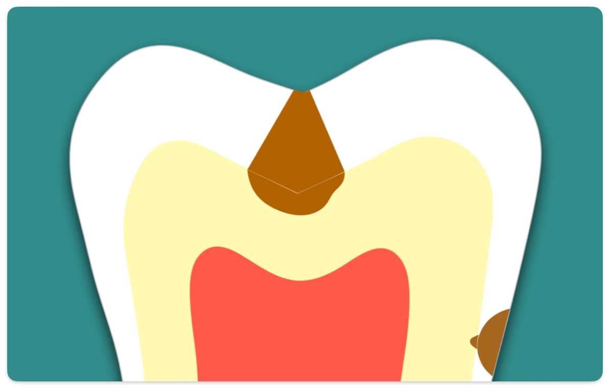
Pit and Fissure caries
- The carious lesion starts along the fissure walls rather than at the base and follows the direction of the enamel rods.
- It forms a triangular or cone-shaped lesion with its apex at outer surface and base toward the DEJ.
- Pit and fissure caries usually produces greater cavitations than smooth surface caries, as the base of the triangle is towards DEJ, it involves greater number of dentinal tubules when the lesion reaches the DEJ.
Zones of Enamel Caries
Most of the histological description of enamel caries is in relation to early lesions. The initial lesion has been divided into four zones based on its histological appearance when longitudinal ground sections are examined with the light microscope.
Starting from inside out, these are : translucent zone, dark zone, body of the lesion and surface layer.
Zone 1: The Translucent Zone
- It is the first recognisable zone of alteration from normal enamel, that lies at the advancing front of the enamel caries and is present only in half of the cases.
- Can be observed when a longitudinal ground section is examined in a clearing agent having a refractive index(RI) identical to that of enamel (eg. Quinoline - RI 1.62).
- Appears structureless, when examined in transmitted light after imbibition with quinoline (The pores are located at prism boundaries and other junctional sites, hence, normal structural markings are not visible when the pores are filled with a medium having the same refractive index as enamel).
- Slightly more porous than sound enamel (pore volume of 1% compared with 0.1% in sound enamel).
- Fluoride content is more relative to sound enamel, with preferential removal of magnesium and carbonate rich mineral.
Zone 2: The Dark Zone
- It lies superficial to the translucent zone and formed as a result of demineralisation.
- It is usually present, hence also referred to as the 'positive zone'.
- Appears dark brown in ground sections examined by transmitted light after imbibition with quinoline. (Since, the micro pores remain filled with air or vapor, light is scattered on passing through the zone, causing brown discolouration of the dark zone).
- Shows positive birefringence (sound enamel -> negative birefringence), when examined with polarising microscope after imbibition with quinoline. (Due to the presence of medium or low refractive index within the micro pore system.)
- Pore volume : 2-4%.
Zone 3: The Body of Lesion
- It is the area of greatest demineralisation, present between the relatively unaffected surface layer and the dark zone.
- Appears relatively translucent compared to sound enamel with well marked striae of Retzius, when examined in quinoline with transmitted light.
- Shows positive birefringence when examined with polarised light after imbibition with water.
- There is reduction in mineral content and increase in unbound water and organic content (due to ingress of bacteria and saliva).
- Pore volume : 5% in spaces near the periphery and 25% in the center of the lesion.
Zone 4: The Surface Zone
- It is well mineralised (partial demineralisation of 1-4%) and remain intact, due to remineralisation i.e., re-precipitation of the calcium and phosphate ions released by subsurface dissolution.
- Eventually, the surface zone is demineralised, usually when the carious process has penetrated some way into the dentin.
- Shows negative birefringence, lying superficial to the positively birefringent body of the lesion, when examined with polarised light after imbibition with water.
- Pore volume : Less than 5%.
Dentinal Caries
Caries of the dentin begins with the natural spread of the disease process along the DEJ and the rapid involvement of great numbers of dentinal tubules, each of which acts as a potential pathway leading to the dental pulp.
Early Dentin Changes
Fatty degeneration of odontoblast process
- Although it is not degenerative process, there is deposition of fat globules.
- It is suggested that fat contributes to the impermeability of the dentinal tubules and may be a predisposing factor in dentinal sclerosis.
Dentinal sclerosis
- There is calcification of the dentinal tubules that tends to seal them off against further penetration by microorganisms.
- Seen prominently in slow chronic cases, while it is minimal in rapidly advancing caries.
- Appears dark in reflected light.
Initial decalcification
- Involves the walls of dentinal tubules, allowing them to distend slightly as they become packed with microorganisms.
Microbial invasion
- Acidogenic organisms predominate early caries as carbohydrate substrate is easily available.
- Proteolytic organisms are predominant in deeper caries due to high protein content of dentin.
Advanced dentinal changes
-
Confluence of the dentinal tubules due to decalcification of their walls.
-
Thickening and swelling of the sheath of Neumann at irregular intervals along the course of involved dentinal tubules.
-
Increase in diameter of dentinal tubules due to packing of tubules by microorganisms.
-
Tiny liquefaction foci
- Ovoid area of destruction filled with necrotic debris that tend to increase in size by expansion.
- Formed by focal coalescence and breakdown of a few dentinal tubules.
- Produces compression and distortion of adjacent dentinal tubules, bending them around the liquefaction focus.
-
Decalcification and proteolysis (matrix destruction) : The process of decalcification (by acidogenic organism) followed by destruction of remaining organic matrix by proteolysis (proteolytic organism) occurs at numerous focal areas. These focal areas eventually coalesce to form a necrotic mass of dentin of leathery consistency.
-
Transverse clefts
- Formed due to extension of the carious process along the lateral branches of the tubules or along the matrix fibres that run at right angles to the dentinal tubules.
- These clefts are parallel to the incremental lines of the dentin and leads to peeling away thin layers with hand instruments during excavation.
Zones of Dentinal Caries
Caries process in the dentin tend to assume the shape of a triangle with the apex toward the pulp and the base toward the enamel.
Starting from pulpal side, at the advancing edge of the lesion, adjacent to the normal dentin, the following zones can be distinguished:
- Zone 1: Zone of fatty degeneration of odontoblast process.
- Zone 2: Zone of dentinal sclerosis characterised by deposition of calcium salts in dentinal tubules.
- Zone 3: Zone of decalcification of dentin.
- Zone 4: Zone of bacterial invasion of decalcified but intact dentin.
- Zone 5: Zone of decomposed dentin.
Also, the zones of dentinal caries (inside out) can be divided as,
- Sound/Normal dentin.
- Sub-transparent dentin: Self-repairable (affected dentin).
- Transparent dentin: Self-repairable (affected dentin).
- Turbid dentin: Dry and leathery, not self-repairable.
- Infected dentin.
Points to Note
- Smooth surface caries appear as an area of decalcification beneath the dental plaque, resembling a smooth chalky surface, while, in pit and fissure caries, chalkiness or yellow, brown or black discolouration may be seen.
- A brown stain in newly erupted teeth is indicative of underlying decay, while, in older individuals it may be due to arrested caries.
- The rate at which caries progress, tends to be slower in older adults because of the generalised dentinal sclerosis that occurs with aging.
- Pioneer bacteria: Microorganisms found penetrating the dentinal tubules in the earliest stages of caries before there is any clinical evidence of the carious process.
- Caries spreads rapidly in malacotic or soft teeth: Composed of considerable amount of interglobular dentin -> decalcification and confluence of tubules occur rapidly in areas of interglobular dentin.
- Secondary dentin involvement: The carious process is slower since the dentinal tubules are fewer in number and more irregular in their course, which delays penetration of the invading microorganisms.
References
- Shafer, Hine, Levy Shafer's Textbook of Oral Pathology (7th Edition), Editors - R Rajendran, B Sivapathasundharam, Elsevier.
*This article is an excerpt from the above mentioned book and Medical Sutras does not make any ownership or affiliation claims.