Gram stain, devised by histologist Christian Gram (1884), differentiates bacteria into two groups:
- Gram-positive: Appears violet (Resist decolorisation and retain the primary stain).
- Gram-negative: Appears red (Discolorized by organic solvent and takes the the secondary stain).
Theories for the difference in staining:
- Gram-positive cells have more acidic protoplasm, hence retain the primary dye more strongly.
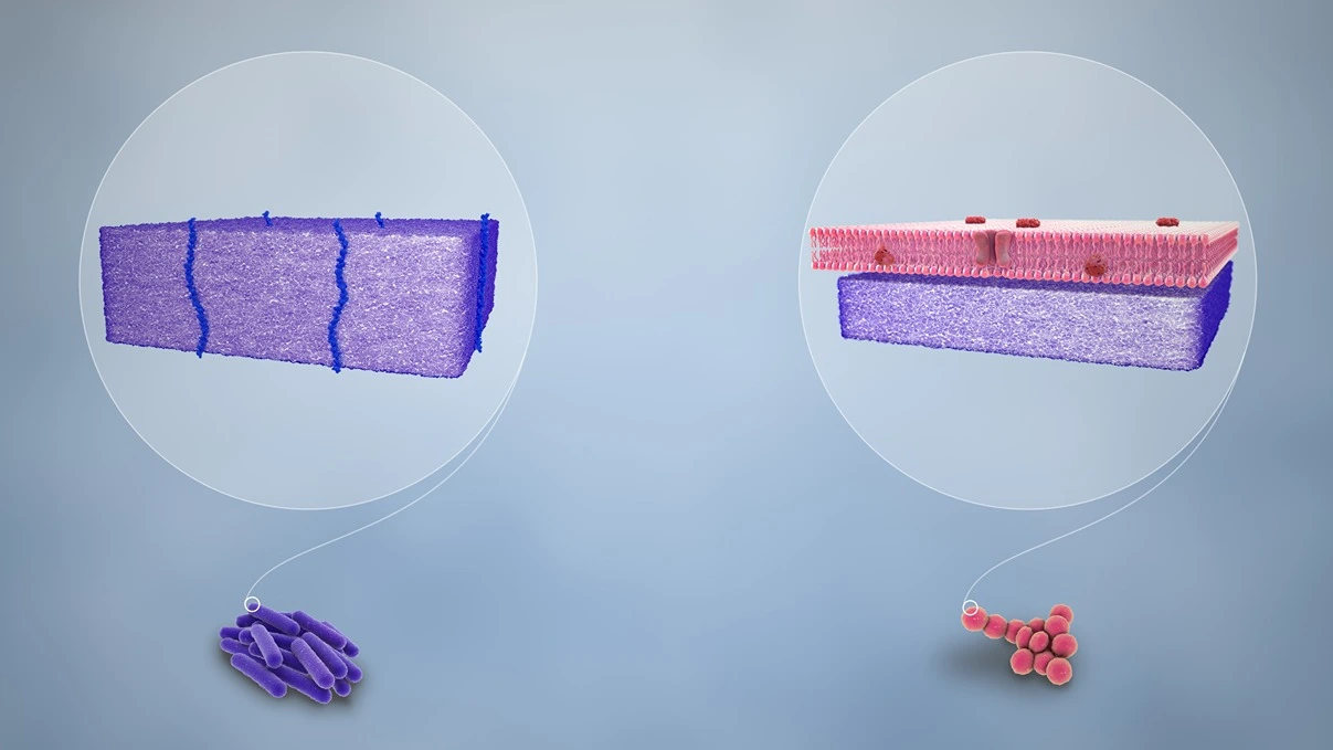
- The cell wall in Gram-positive cells is entirely made up of thick peptidoglycan layer (15-80 nm), thus retains the dye-iodine complex.
- The high lipid content of Gram-negative bacteria makes them permeable to secondary dye after decolorisation with organic solvents.
Method of Gram Staining
- Primary staining is done with a para-rosaniline dye such as crystal violet, methyl violet or gentian violet.
- A dilute solution of iodine is applied, so that primary dye-iodine complex is formed.
- Decolorisation with an organic solvent such as ethanol, acetone or aniline.
- Counterstaining with a secondary dye of contrasting color such as carbol fuchsin, safranine or neutral red.
Gram-positive Bacilli
-
Corynebacteria.
-
Bacillus anthracis (Gram-variable, tends to be decolorised easily).
-
Clostridium.
-
Eubacterium.
-
Propionibacterium.
-
Lactobacillus.
-
Mobiluncus.
-
Actinomycetes (filamentous)
- Anaerobic : Actinomyces, Arachnia, Bifidobacterium, Rothia.
- Aerobic : Nocardia, Actinomadura, Dermatophilus, Streptomyces.
Gram-positive Cocci
- Staphylococci.
- Streptococci.
- Peptostreptococcus.
- Peptococcus.
Gram-negative Bacilli
-
Bacteroides.
-
Prevotella.
-
Porphyromonas.
-
Fusobacterium.
-
Leptotrichia.
-
Enterobacteriaceae :
- Escherichia coli, Shigella.
- Salmonella.
- Citrobacter.
- Klebsiella, Enterobacter.
- Proteus.
- Yersinia.
-
Vibrio cholerae.
-
Pseudomonas.
-
Haemophilus.
-
Pasteurella.
-
Francisella.
-
Bordetella.
-
Brucella.
-
Listeria monocytogenes.
-
Helicobacter pylori (spiral rod).
-
Rickettsia, Orientia and Ehrlichia.
-
Bartonella.
Gram-negative Cocci
- Nisseria.
- Veillonella.
- Moraxella catarrhalis.
- Coxiella burnetii (cocobacillus)
Points to Note
- Gram-positive and gram-negative bacterias differ significantly in several properties such as structure, growth requirements, pathogenicity and susceptibility to antibiotics.
- Decolorisation is not full proof, as inadequate decolorisation may cause all cells to appear gram-positive and even gram-positive cells may be decolorised on prolonged treatment with the organic solvent.
- Gram-positive bacteria may become gram-negative when their cell wall is damaged.
References
-
Ananthanarayan and Paniker's Textbook of Microbiology, 10th Edition (Editor - Reba Kanungo), Universities Press.
-
The image used is licensed under the Creative Commons Attribution-Share Alike 4.0 International license.
- Description : Gram-positive bacteria(left) with thick peptidoglycan layer stains.
- Source : http://www.scientificanimations.com/wiki-images/.
- Author : https://www.scientificanimations.com/.
*This article is an excerpt from the above mentioned book and Medical Sutras does not make any ownership or affiliation claims.