It is the ninth cranial nerve, that is composed of both the motor and sensory fibres (mixed nerve).
It provides sensory innervation to the tongue and pharynx, hence, known as glossopharyngeal nerve.
Functional Components
Special Visceral Efferent Fibres
- Arise from nucleus ambiguus.
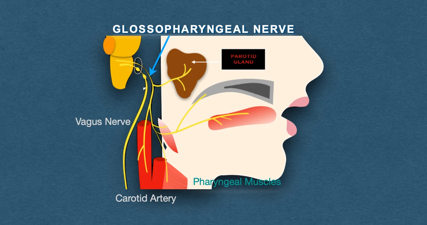
- Supplies the stylopharyngeus muscle.
General Visceral Efferent Fibres
- Are preganglionic parasympathetic fibres, that arise from the inferior salivatory nucleus.
- Supplies the secretomotor fibres to the parotid gland.
Special Visceral Afferent FIbres
- Carry taste sensations from the posterior 1/3rd of tongue including vallate papillae.
- Terminate in the dorsal nucleus of the vagus.
General Somatic Afferent Fibres
- Carry proprioceptive sensations from the stylopharyngeus and skin of the auricle.
- Terminate in the nucleus of the spinal tract of the trigeminal nerve.
Anatomy & Pathway
It arises from the upper part of the lateral aspect of the medulla between the olive and the inferior cerebellar peduncle by 3-4 rootlets.
- The rootlets unite to form a single trunk which runs forward and laterally, passes through the intermediate compartment of the jugular foramen and leaves the cranial cavity. In the jugular foramen, the superior and inferior sensory ganglia are located.
- After exiting the cranial cavity, the nerve passes downward and forward between the internal carotid artery and the internal jugular vein.
- It then descends anterior to the internal carotid artery to the styloid process and muscles attached to it and reaches the lower border of the stylopharyngeus.
- It passes along with the stylopharyngeus through the gap between the superior and middle constrictors of the pharynx, and curves forward along the lateral aspect of the stylopharyngeus muscle.
- Then, it passes deep to the stylohyoid ligament and posterior edge of the hyoglossus muscle, where it breaks into terminal branches.
Branches & Distribution
Tympanic Branch (Jacobson's Nerve)
- Leaves the inferior ganglion and enters the middle ear through the tympanic canaliculus.
- Forms the tympanic plexus over the promontory of the middle ear.
- Branches of tympanic plexus: Lesser petrosal nerve, and twigs to tympanic cavity, auditory tube, and mastoid air cells.
- Lesser petrosal nerve: Carries preganglionic parasympathetic fibres that relay in the otic ganglion and the postganglionic fibres from the ganglion supply the parotid gland.
Carotid Nerve (Nerve of Herring)
- It is a branch to carotid sinus and carotid body.
- Serves as an afferent limb for pressoreceptor and chemoreceptor reflexes from the carotid sinus and carotid body to regulate the heart rate and respiration, respectively.
Pharyngeal Branch
- Joins the pharyngeal branches of the vagus and the cervical sympathetic chain to form the pharyngeal plexus on the middle constrictor of the pharynx.
Branch to Stylopharyngeus
- Only motor branch of the glossopharyngeal nerve.
- Arises as the nerve, and, winds round the stylopharyngeus muscle.
Tonsillar Branches
- Supply the mucous membrane of tonsil, fauces and palate.
Lingual Branches
- Supply the posterior 1/3rd of the tongue and vallate papillae.
- Convey taste and general sensations.
Clinical Significance
A complete lesion of glossopharyngeal nerve results in:
- Loss of taste and general sensations over the posterior one-third of the tongue.
- Difficulty in swallowing.
- Loss of salivation from the parotid gland.
- Unilateral loss of the gag reflex.
*Isolated lesion of glossopharyngeal nerve is rare and vagus nerve is often involved.
Glossopharyngeal Neuralgia
- Rare, but may occur.
- Characterised by paroxysmal attacks of intractable pain in the area of the sensory distribution of the glossopharyngeal nerve such as throat, tongue and ear, precipitated by swallowing.
Clinical Testing
- Eliciting the gag reflex: Tickling the posterior wall of the pharynx, soft palate, or tonsillar fossa, leads to reflex contraction of pharyngeal muscles, resulting in gagging and retching.
- Testing taste sensations in the posterior one-third of the tongue.
References
| [](https://amzn.to/3Ixnict)
Textbook of Anatomy Head, Neck, and Brain (Volume III), Vishram Singh |
- The image used is licensed under the Creative Commons Attribution 4.0 International license. (Source: Romano, N., Federici, M. & Castaldi A. Imaging of cranial nerves: a pictorial overview. Insights Imaging 10, 33 (2019). https://doi.org/10.1186/s13244-019-0719-5.)
*This article is an excerpt from the above mentioned sources and Medical Sutras does not make any ownership or affiliation claims.