Gingival cyst of adult is a small, uncommon developmental odontogenic cyst of the gingival soft tissue (free or attached gingiva) that contains a lining epithelium of cuboidal cells and distinctive focal thickenings.
Origin
The possible sources of gingival cyst includes:
- Cystic transformation of the remnants of dental lamina or the glands or rests of Serres.
- Traumatic implantation of surface epithelium (not truly odontogenic cyst).
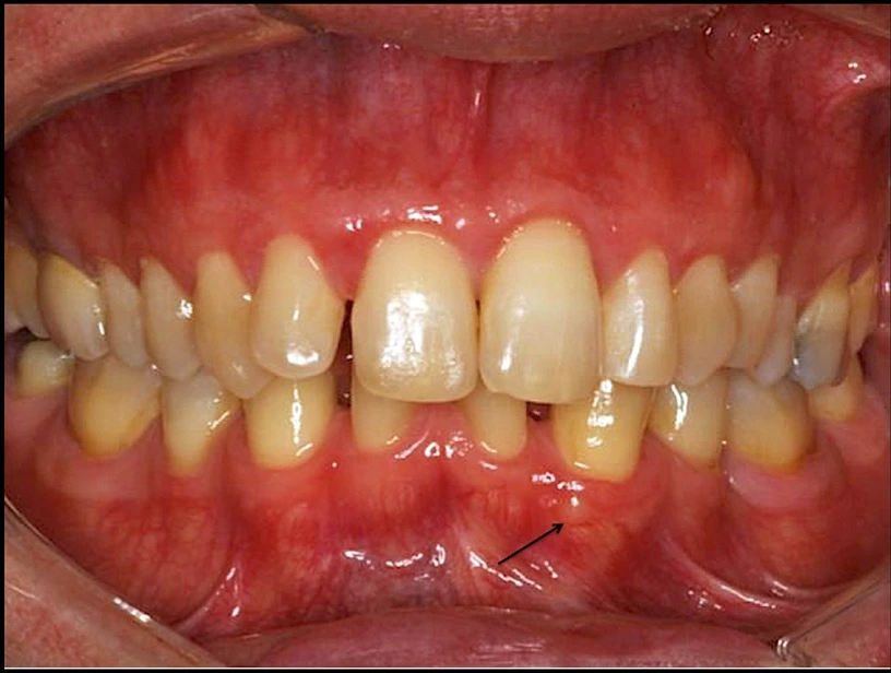
Clinical Findings
- Small, well-circumscribed, painless swelling of the gingiva.
- More common in mandibular premolar and canine region, with predilection for males.
- The lesion resembles adjacent mucosa or bluish in colour.
- Size : Less than 1 cm in diameter.
- The lesions are soft and fluctuant and the adjacent teeth are usually vital.
Histologic Features
- Lined by thin stratified squamous epithelium, ranging from one flattened cell to several cells in thickness.
- Glycogen-rich clear cells may be present, especially in the focal thickenings or plaques of the lining.
- Dental lamina rests may also be found in the connective tissue wall and are commonly composed of the same type of glycogen-rich clear cells.
- May be unicystic or polycystic.
Treatment
Local surgical excision is usually recommended and the lesions do not tend to recur.
Points to Note
- Wysocki and colleagues concluded that gingival cyst arise from postfunctional rests of dental lamina and represents the extraosseous counterpart of lateral periodontal cyst.
- GIngival cyst is a true cyst as it consists of a pathologic epithelium lined cavity that usually contains fluid.
Dental Lamina / Gingival Cyst of Newborn
- Dental lamina cyst of the newborn comprise of multiple, occasionally solitary, superficial raised nodules on edentulous alveolar ridges of infants.
- They are derived from rests of the dental lamina and consist of keratin-producing epithelial lining.
- Whitish in colour, as they are filled with keratin.
- Histologically, they present with a thin epithelial lining which lacks rete processes and a lumen usually filled with desquamated keratin, occasionally containing inflammatory cells.
- No treatment is required as they usually disappear by opening onto the mucosal surface or through disruption by erupting teeth.
Epstein's Pearls
- Cystic, keratin-filled nodules found along the mid-palatine raphe, probably derived from entrapped epithelial remnants along the line of fusion.
Bohn's Nodules
- Keratin-filled cysts scattered over the palate, most numerous along the junction of the hard and soft palate and apparently derived from the palatal salivary gland structures.
References
-
Shafer, Hine, Levy Shafer's Textbook of Oral Pathology (7th edition), Editors - R Rajendran, B Sivapathasundharam, Elsevier.
-
Textbook of Oral Medicine (3rd edition), Anil Govindrao Ghom, Savita Anil Ghom (Lodam), Jaypee Brothers Medical Publishers (P) Ltd.
-
The image used is licensed under the Creative Commons Attribution-Share Alike 4.0 International license.
- Source : Gingival Cyst of the Adult: Report of an Inconspicuous Lesion Associated with Multiple Agenesis. Case Reports in Dentistry Volume 2017 (2017), Article ID 4346130, 4 pages. https://doi.org/10.1155/2017/4346130.
- Author : Juliana Mançano Melhado Brod, Fabrício Passador-Santos, Andresa Borges Soares, and Marcelo Sperandio.
*This article is an excerpt from the above mentioned books and Medical Sutras does not make any ownership or affiliation claims.