It is the 7th cranial nerve, that consist of both motor and sensory components (mixed nerve).
-
It supplies the muscles of facial expression, hence, called the facial nerve.
-
It consists of two roots:
- Motor root (facial nerve proper): Larger and medial.
- Sensory root (nervous intermedius): Small and located laterally.
-
It is the most frequently paralysed of all the peripheral nerves of the body.
Functional Components
Special Visceral Efferent Fibres
- Arise from the motor nucleus of the facial nerve in the pons.
- Supply the muscles of facial expression.
General Visceral Efferent Fibres
- Preganglionic parasympathetic fibres.
- Arise from the lacrimatory and salivatory nuclei in the brainstem.
- Supply the secretomotor fibres to lacrimal, submandibular, and sublingual glands.
Special Visceral Afferent Fibres
- Carries special sensations of taste from anterior 2/3rd of the tongue except vallate papillae.
- Terminate in the nucleus of tractus solitarius (gustatory nucleus) in the brainstem.
- The cell bodies of SVA and GVA fibres are located in the geniculate ganglion.
General Somatic Afferent Fibres
- Carries general sensations from the skin of the auricle.
- Terminate in the spinal nucleus of the trigeminal nerve.
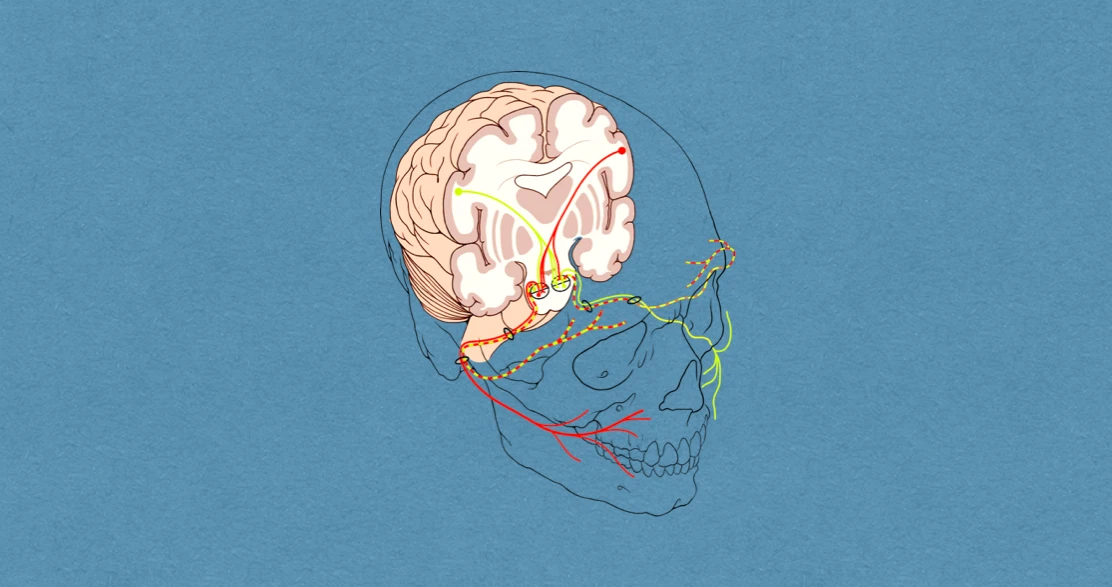
Anatomy & Pathway
Origin
Arises from the pontomedullary junction lateral to the superior end of the olive of the medulla. The sensory root lies between the motor root (medially) and the vestibulocochlear nerve (laterally).
Course
- After emerging the brainstem, the roots of the facial nerve pass laterally and forward in the cerebellopontine angle, along with the vestibulocochlear and labyrinthine artery.
- These structures then enter the internal acoustic meatus.
- In the meatus, the motor root is lodged in a groove on the vestibulocochlear nerve, while the sensory root remains separate.
- At the lateral end (bottom) of the internal acoustic meatus, the two roots unite to form the trunk of the facial nerve.
- The nerve enters the facial canal in the petrous temporal bone through its opening in the fundus of the internal acoustic meatus.
- The facial canal divide into three segments: labyrinthine, tympanic, and mastoid.
- The facial nerve comes out of the cranial cavity through the stylomastoid foramen.
Branches & Distribution
Greater Petrosal Nerve
- Arises from the geniculate ganglion.
- Consists of preganglionic parasympathetic fibres that relay in the pterygopalatine ganglion.
- Supplies the secretomotor fibres to the lacrimal gland and mucous glands of nasal cavity and palate.
Nerve to Stapedius
- Arises from the vertical part of the facial nerve opposite the pyramidal eminence.
- Supplies the stapedius muscle.
Chorda Tympani Nerve
-
Arises from the vertical part of the facial nerve about 6 mm above the stylomastoid foramen.
-
Consists of two types of fibres:
- Preganglionic parasympathetic fibres (GVE): Provides secretomotor supply to the submandibular and sublingual glands.
- Special visceral afferent fibres (SVE): Carries taste sensations from anterior 2/3rd of the tongue.
-
It enters the middle ear through the posterior canaliculus, runs across its lateral wall, passing between the long process of incus and the handle of malleus, and leaves the middle ear by entering the anterior canaliculus.
-
Then, the nerve enters the infratemporal fossa through the medial end of petrotympanic fissure.
-
It runs medially forward and downward, crossing the medial aspect of the spine of sphenoid, to join the posterior aspect of the lingual nerve.
Posterior Auricular Nerve
- Supplies the occipital belly of occipitofrontalis.
Nerve to Posterior Belly of Digastric
- Supplies the posterior belly of digastric.
Nerve to Stylohyoid
- Supplies the stylohyoid muscle.
Terminal Branches
- Temporal
- Zygomatic
- Buccal
- Marginal mandibular
- Cervical
Clinical Significance
Supranuclear Lesion
- Paralysis of only lower half of the opposite side of face.
- Spares the upper part of face, since, it is innervated by the corticonuclear fibres of both the cerebral hemispheres.
Infranuclear Lesion
Involve whole of the same side of face, with signs and symptoms differing according to the side of the lesion:
- At or just above the stylomastoid foramen: Causes Bell's palsy, that involves loss of motor functions of all muscles of facial expression. There is deviation of mouth toward the normal side, inability to close the mouth and eye, and, accumulation of food in the vestibule of mouth.
- Above the origin of Chorda tympani: All above signs and symptoms, with decreased salivation and loss of taste sensations in the anterior 2/3rd of the tongue.
- Above the origin of nerve to stapedius: In addition to above, there is enhanced sensitivity to hearing (hyperacusis).
- At the geniculate ganglion: Loss of lacrimation, in addition to all of the above signs and symptoms.
Crocodile Tears Syndrome
- Characterised by paroxysmal lacrimation during eating.
- Occurs in case of facial nerve lesion proximal to the geniculate ganglion.
- Here, the preganglionic fibres meant to provide secretomotor supply to the submandibular and sublingual glands are misdirected and grow in the endoneurial sheaths of preganglionic secretomotor fibres supplying the lacrimal gland.
Ramsay Hunt Syndrome
-
Occurs due to involvement of geniculate ganglion in herpes zoster infection.
-
It presents clinically with:
- Herpetic vesicles on the auricle.
- Hyperacusis.
- Loss of lacrimation.
- Loss of taste sensations in the anterior 2/3rd of tongue.
- Complete ipsilateral facial palsy.
References
| [](https://amzn.to/3Ixnict)
Textbook of Anatomy Head, Neck, and Brain (Volume III), Vishram Singh |
- The image used is licensed under the Creative Commons Attribution 2.5 Generic license. (Author: Patrick J. Lynch, medical illustrator. http://patricklynch.net)
*This article is an excerpt from the above mentioned sources and Medical Sutras does not make any ownership or affiliation claims.