Acccording to the Glossary of Endodontic Terms, caries is defined as a localised and progressive bacterial infection that results in disintegration of the tooth, usually beginning with the demineralisation of enamel and followed by bacterial invasion.
Superficial caries needs to be differentiated from Enamel hypocalcificaiton, Enamel hypoplasia, and Dental fluorosis.
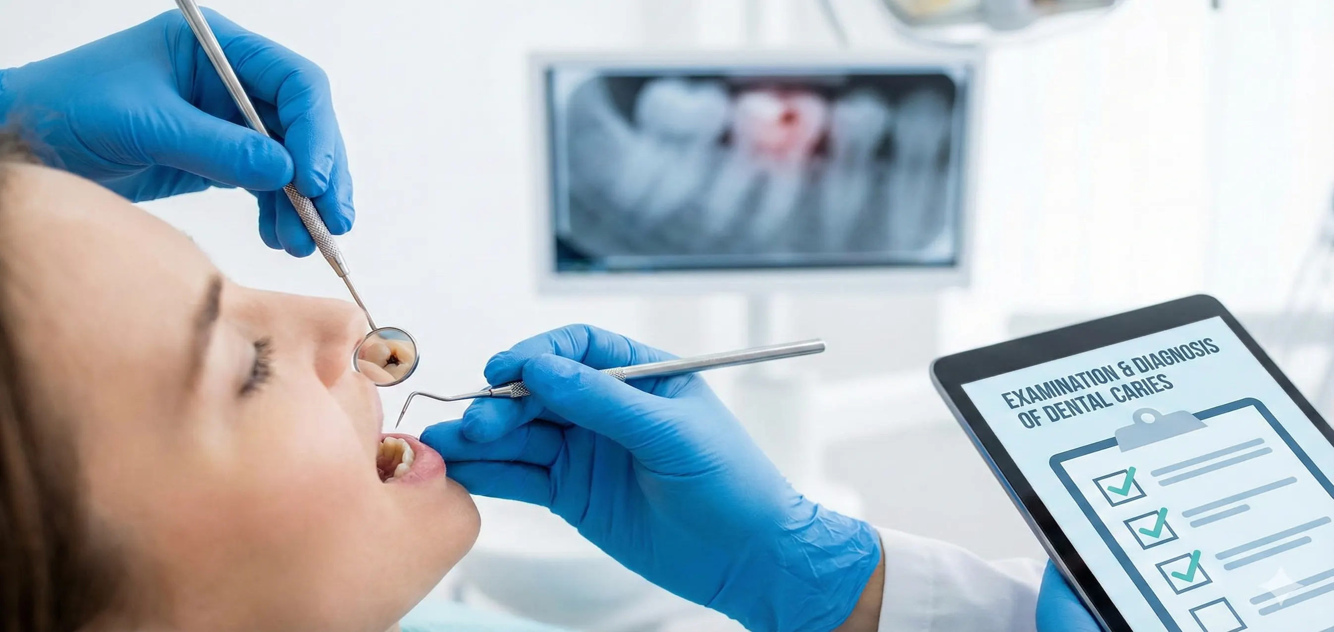
There are several methods for examination and diagnosis of dental caries. These include:
- Conventional diagnostic methods: Inspection, Probing and Percussion.
- Special diagnostic methods: Radiographs, Cold and heat test, Dental floss examination, and Diagnostic cavity preparation.
- New technologies: Fiber-optic transillumination, Electrical impedance, Ultrasonics, Elastomeric separating modulus, Staining, and Quantitative laser fluorescence.
Conventional Diagnosis Methods
Inspection
- It involves obersving the tooth surface and other areas using a mouth mirror.
- Carious teeth may present as black or chalky areas or a formed cavity.
Probing
- It involves using a sharp probe (dental explorer) to inspect the suspicious areas and examine the depth and extension of carious lesion.
- It can help to locate affected areas, areas of dentinal hypersensitiviy and pulp exposure in deep caries.
- Probing is particularly useful in cases where a proximal caity is suspected and cannot be located through visual inspection. The explorer tip gets hooked by the edge of the cavity, thus helping in locating the cavity.
Percussion
- Caries does not cause periodontal and periapical inflammation, so the reaction to percussion is always negative.
- This method is helping in identifying carious lesion extending periapically. For example, acute apical periodontitis presents a painful response to percussion.
Special diagnostic methods
Radiographic Examination
-
Carious lesions presents as radiolucency on the hard dental tissues (enamel and dentin), due to demineralisation.
-
Radiographs are helpful in locating proximal caries, subsurface caries and secondary caries. They can also be used to assess the proximity of caries to pulp chamber.
-
The commonly used x-rays include periapical and bite-wing x-rays.
-
The radiographic findings must be correlated with clinical examination, since, radiographs give a two-dimensional image and there may be some overlap with other radiolucent areas.
-
Important differentials to be considered include:
- Proximal caries and normal triangular low-density areas in the cervical region of the tooth.
- Secondary caries and low-density base materials used on the floor of cavity.
Cold and Heat Irritation Test
-
It involves assessing the response of dental pulp to cold and hot stimuli. These external stimuli elicit acute pain and helps to determine the severity of caries.
-
The test includes placing chloroethane-soaked cotton ball or hot gutta percha stick on the tooth surface and evaluating the patient's response.
- Healthy pulp: Mild-to-moderate sharp pain that subsides immediately (within 1–2 seconds) after the stimulus is removed.
- Reversible pulpitis (Exaggerated but fast): A sharp, quick response that disappears quickly.
- Irreversible pulpitis (Lingering pain): The pain persists for several seconds or minutes after the stimulus is gone. If the pain becomes spontaneous (happening without any stimulus), the diagnosis almost always shifts toward irreversible damage.
- Pulp necrosis (No response): The patient feels nothing, although false negatives can occur in teeth with receded pulps or recent trauma.
-
Using cold water as a stimulus is less precise than a cotton pellet and is not recommended: Water can flow across multiple teeth or deep into a cavity, making it difficult to pinpoint exactly which tooth or which part of the tooth is triggering the pain.
-
Caries vs Dentin Hypersentivity
- In caries, pain is usually triggered by chemical (sugar) or thermal stimuli and is localized to the site of decay where the enamel is compromised.
- Dentin hypersensitivity is typically characterized by a short, sharp and quickly reversible pain arising from exposed dentin. It is a response to stimuli that would not normally cause pain in a healthy tooth, but unlike pulpitis, it never lingers.
Dental Floss Examination
- This method is useful in indentifying caries in proximal areas that are difficult to examine by inspection and probing.
- It involves putting a dental floss across the embrasure of the suspicious tooth surface and movinf the floss horizontally in a seesaw motion. This way the examiner can experience the roughness of the surface. The floss will be torn if caries is present.
- However, presence of calculus can mislead the examination with floss.
Diagnostic Cavity Preparation
- It involves removal of supportless enamel and infectious tissue on the floor and wall of the cavity.
- This helps the examiner to clearly visualise and define the affected area and pulp condition.
- Also, the decalcified dentin can be stained with 0.5% basic fuschin, for better identification and removal of infectious dentin.
New technologies
Fiber-optic transillumination
Electrical impedance
Ultrasonics
Elastomeric separating modulus
Staining
Quantitative laser fluorescence