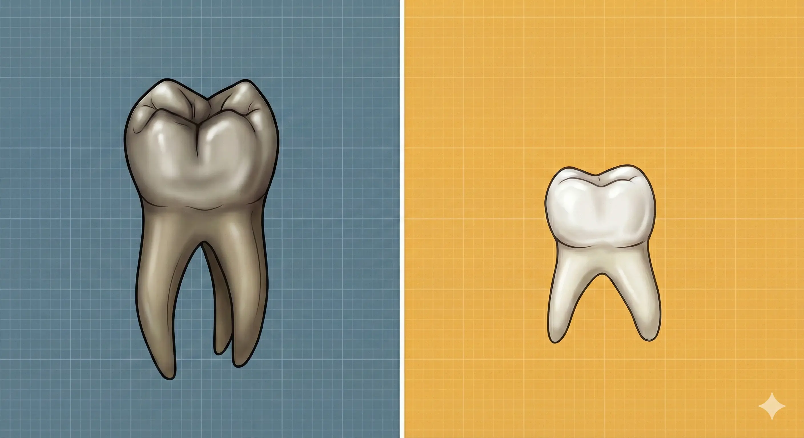
The primary teeth are smaller in overall size and crown dimensions when compared to their counterparts in the permanent dentition. The primary teeth are lighter (whiter) in appearance as they are usually less pigmented.
The key differences between primary and permanent teeth are summarised below:
Primary Anterior Teeth
- Crowns are wider mesiodistally in comparison with their crown length.
- Roots are narrower and longer comparatively.
- The cervical ridges of enamel are more prominent.
Primary Posterior Teeth
- The roots are longer, more slender and flared, extending out beyond projected outlines of the crowns. This flare allows more room between the roots for the development of permanent tooth crowns.
- The crowns and roots are more slender mesiodistally at their cervical portions.
- The buccolingual diameter of primary molars is less than that of permanent teeth.
- The cervical ridges on the primary molars are much more pronounced, especially on the buccal aspect of the first molars.
- The buccal and lingual surfaces of primary molars are flatter above the cervical curvatures, which narrows the occlusal surfaces.
Cross-sectional Analysis
- Crown widths in all directions are large compared with root trunks and cervices.
- The enamel is relatively thin and has a more consistent depth.
- Enamel rods at the cervix slope occlusally, instead of gingivally as in the permanent teeth.
- Dentin thickness between the pulp chambers and the enamel is limited, particularly in some areas (lower second primary molar).
- Dentin thickness is comparatively greater over the pulpal wall at the occlusal fossa of primary molars.
- The primary teeth have large pulp chambers with higher pulp horns.
- Roots are narrow and long compared with the width and length of the crown.
- Roots of molars flare markedly and thin out rapidly as they approach the apices.
References
- Wheeler's Dental Anatomy, Physiology and Occlusion (2019), Stanley J. Nelson DDS MS, Elsevier.
*This article is excerpt from the above mentioned book and Medical Sutras does not make any ownership and affiliation claims.