Defined as an odontogenic cyst that surrounds the crown of an impacted tooth and is caused by the accumulation of fluid between the reduced enamel epithelium and the enamel surface.
- Most common type of developmental odontogenic cyst, estimated to be about 20% of all jaw cysts.
Clinical Features
-
Usually solitary and associated with the crown of an impacted, embedded or unerupted tooth; may also be found with complex compound odontoma or supernumerary tooth.
-
Common sites : Mandibular and maxillary third molar and maxillary cuspid regions.
-
Associated syndromes : Cleidocranial dysplasia, Maroteaux-Lamy Syndrome.
-
The dentigerous cyst is potentially capable of becoming an aggressive lesion. Possible sequelae associated with the continued enlargement of the cyst are:
- Facial asymmetry.
- Extreme displacement of teeth.
- Severe root resorption.
- Pain.
Radiographic Features
-
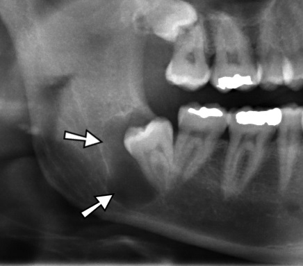
Appears as a radiolucent area surrounding the crown of unerupted or impacted tooth.
-
It can be differentiated from normal follicular space (3-4 mm), when the size of radiolucency is more than 5 mm.
-
Radiologic variations :
- Central : Crown is enveloped symmetrically.
- Lateral : Cyst is located on one side of the crown.
- Circumferential : Entire tooth appears to be enveloped by the cyst.
Histologic Features
- Nothing characteristic to differentiate the dentigerous cyst from other odontogenic cysts.
- Histologic examination shows a thin connective tissue wall with a thin layer of stratified squamous epithelium lining the lumen.
- Islands of odontogenic epithelium : seen in both normal dental follice and dentigerous cyst.
- Rete peg formation : seen in case of secondary infection.
- Rushton Bodies : Peculiar, linear, hyaline bodies with variable stainability found within the lining epithelium, in case of inflammation.
- Cystic fluid : Thin, watery yellow, occasionally blood tinged.
Treatment
- Dictated by the size of the lesion.
- Small cysts can be surgically removed entirely.
- Larger cysts that involves extensive boss loss, require marsupialization or inserstion of a surgical drain.
Potential Complications
- Ameloblastoma : can develop from the lining epithelium or from rests of odontogenic epithelium present in the cystic wall.
- Epidermoid carcinoma : can develop from the lining epithelium or from rests of odontogenic epithelium present in the cystic wall.
- Mucoepidermoid carcinoma : can develop from the lining epithelium that contains mucus-secreting cells.
References
-
Shafer, Hine, Levy Shafer's Textbook of Oral Pathology (7th Edition), Editors - R Rajendran, B Sivapathasundharam, Elsevier.
-
The image used is licensed under the Creative Commons Attribution 3.0 Unported License.
- Description : Dentigerous jaw cyst in the right mandible around and impacted wisdom tooth.
- Source : Derived from JawCyst.jpg.
- Author : Coronation Dental Specialty Group, derivative work Jbarta and Mikael Haggstrom.
*This article is an excerpt from the above mentioned books and Medical Sutras does not make any ownership or affiliation claims.