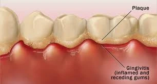
Plaque is the soft, non-mineralised, bacterial deposit that forms on surfaces of inadequately cleaned teeth and dental prostheses and resists removal by physiologic and oral cleansing forces such as saliva and tongue movement (removable by toothbrushing).
Plaques can be classified based on the anatomical area in which they form:
- Supragingival plaque: Plays important role in the pathogenesis of dental caries.
- Subgingival plaque: Responsible for the initiation of periodontal diseases.
Composition
Plaque is composed of about 80% water and 20% solids.
- Salivary components: Mucin, desquamated epithelial cells and microorganisms.
- Bacterial and salivary proteins: 50% of the dry weight.
- Carbohydrates and Lipids (25% of the dry weight): Most of the carbohydrates in the matrix consist of polymers, glucans, fructans, and heterosaccharides synthesised by the bacteria. Some of these polymers are thought to play a role in bacterial attachment and cohesion, and others are more important as a reservoir of fermentable substrates metabolised by bacteria when other more readily utilised carbohydrates in plaque become depleted.
- Inorganic compounds (5-10% of dry weight): The concentration of calcium and phosphate in dental plaque is several magnitudes higher than in saliva, probably due to the infiltration of salivary proteins containing these constituents in the bound form. Most of the calcium found in plaque is non-ionic and solubilisation occurs as pH drops.
Microbiology of Dental Plaque
The dental plaque predominantly contains three basic groups of microorganisms. Of these, S. mutans is considered to be the chief etiologic agent in human dental caries.
- Streptococci: S. mutans, S. sanguis, S. mitior, S. milleri and S. salivarius (uncommon).
- Actinomyces: A. viscosus, A. naeslundii, A. israelii and Rothia dentocariosa.
- Veillonella: V. parvula and V. alcalescens.
Mechanism of Plaque Formation
Formation of dental plaque requires two types of specific bacterial adherent interactions.
- Firstly, bacteria attach selectively to the acquired pellicle.
- Secondly, bacteria accumulate via specific adhesive and cohesive interactions involving components of the plaque matrix and direct bacterial cell contact.
Acquired Pellicle
- A glycoprotein derived from the saliva that is adsorbed on the tooth surface.
- It is an important component of dental plaque, as it forms just prior to, or, concomitantly with bacterial colonisation and may serve as a nutrient for plaque microorganisms, thus, facilitating plaque formation.
The pellicle appears as three distinct components.
- Subsurface component: Below the enamel surface, have a dendritic configuration.
- Surface component: Closely associated with the tooth surface (Thickness: 1 micrometer).
- Suprasurface component: Has a scalloped appearance (Thickness: 10 micrometer).
Bacterial Adherence
Almost all bacteria and all natural surfaces, including teeth have a net negative charge. Bacteria appear to possess surface components that have recognition potential, which bind to specific receptors on the pellicle and other host tissues. These surface components are referred to as adhesins.
First Phase
- The organisms are thought to be attracted on to the surface by van der Waals forces.
- They are loosely associated as firm contact does not occur because of the repulsive effects of the negative electrostatic charges.
Second Phase
- Appears to involve polymeric substances on the surface of the bacterium which links the organisms to the target surface.
- The polymeric material may bind to the surface by the formation of hydrogen, hydrophobic, ionic or other types of bonds, resulting in a firmer attachment.
Bacterial Accumulation
Both bacterially-derived polymers and salivary components appear to play important roles in this process.
- S. mutans synthesised extracellular glucans and fructans from sucrose but not from other common carbohydrates and this polymer synthesis enables the organism to accumulate in large masses.
- Most tooth-associated streptococci, actinomyces and neisseria can produce extracellular polymer glucan.
- Recent studies have suggested that S. mutans can adsorb on to hydroxyapatite without the synthesis of extracellular polymers and certain S. mutans serotypes can form plaque in the absence of sucrose. However, such plaques are less tenacious to enamel.
Role of Plaque in Dental Caries
When plaque contains appreciable proportions of highly acidogenic bacteria such as S. mutans and is exposed to readily fermentable dietary sucrose, the bacterias produce sufficient concentration of acids to demineralise the enamel.
- Stephan (1940) showed that the pH of plaques varied among different individuals, but averaged about 7.1 in caries-free persons and about 5.5 in persons with extreme caries activity.
- A drop in local pH below 5.5 causes demineralisation of tooth. At a critical pH of 5.5, the tooth minerals act as buffers and they lose calcium and phosphate ions into the plaque. This type of buffering activity would help in maintaining the local pH at about 5.5.
- However, when the local pH falls below 5.0, subsurface demineralisation in inevitable and this results in formation of incipient caries.
- When the pH is lowered further it leads to the surface demineralisation of enamel.
Points to Note
- Materia alba: Loosely adherent whitish material composed of dead cells, food debris and microorganisms, that can be removed mechanically using gauze, water jet or an irrigator.
- Sordes: Whitish gummy material, often present in smaller quantities, and more prominent in fevers.
- Dental calculus: There is mineralisation of both the plaque matrix and the microorganisms. However, the free surface of calculus usually harbors living bacteria.
- Statherin: A salivary protein which maintains super saturation of the fluid phase of plaque with apatite, by adsorbing onto early crystal nuclei and preventing crystal growth.
- Lectins: Protein adhesins that bind to specific sugars.
References
- Shafer, Hine, Levy Shafer's Textbook of Oral Pathology (7th Edition), Editors - R Rajendran, B Sivapathasundharam, Elsevier.
- The image used is licensed under the Creative Commons Attribution-Share Alike 4.0 International license. (Author : Apple Dental, Source : Wikimedia commons).
*This article is an excerpt from the above mentioned book and Medical Sutras does not make any ownership or affiliation claims.