A compound microscope uses more than one lens, i.e., it consists of a series of lenses (standard 2 lens) in a line, where each lens magnifies the image over the previous one.
- Objective lens : Closest to the object or specimen. Consists of 3 or 4 lenses with magnifying power between 40x to 1000x.
- Eyepiece lens : Closest to the observer's eye.
Working Principle
As light through the slide reaches the objective lens, it forms a magnified image, also called as real image. Now, as the light passses through the eyepirce lens, it further magnifies the image and creates the enlarged virtual image seen to the eye.
Parts
Eye lens
- At the upper end of the microscope.
- Magnifies the already magnified image projected by the objective lens before the image reaches the observer.
- Usually 10x but also comes in 5x, 12.5x, 15x and 20x.
Diopter adjustment
- To change focus on one eyepiece so as to correct for any difference in vision between the two eyes.
Body tube (Head)
- Connects the eyepiece to the objective lenses.
Arm
- Connects body tube to base of the microscope.
Coarse adjustment
- Brings the specimen into general focus.
Fine adjustment
- Fine tunes the focus and increases the detail of the specimen.
Nosepiece
- A rotating turret that houses the objective lenses.
Objective lens
- Located on the nosepiece, it is used in combination with the eyepiece to provide a range of magnification from 40x to 100x.
Specimen or slide
- The specimen is the object being examined.
Stage
- Platform for slide.
Stage clips
- Metal clips that hold the slide in place.
Stage height adjustment (Stage control)
- Knobs that move the stage left and right or up and down.
Aperture
- Hole in the middle of the stage that allows light from the illuminator to reach the specimen.
Illumination
- The light source for a microscope.
Diaphragm
- Adjusts the amount of light that reaches the specimen.
Condenser
- Gathers and focuses a cone of light on the slide.
Base
- Supports the microscope and it's where illuminator is located.
References
- Ananthanarayan and Paniker's Textbook of Microbiology, 10th Edition (Editor - Reba Kanungo), Universities Press.
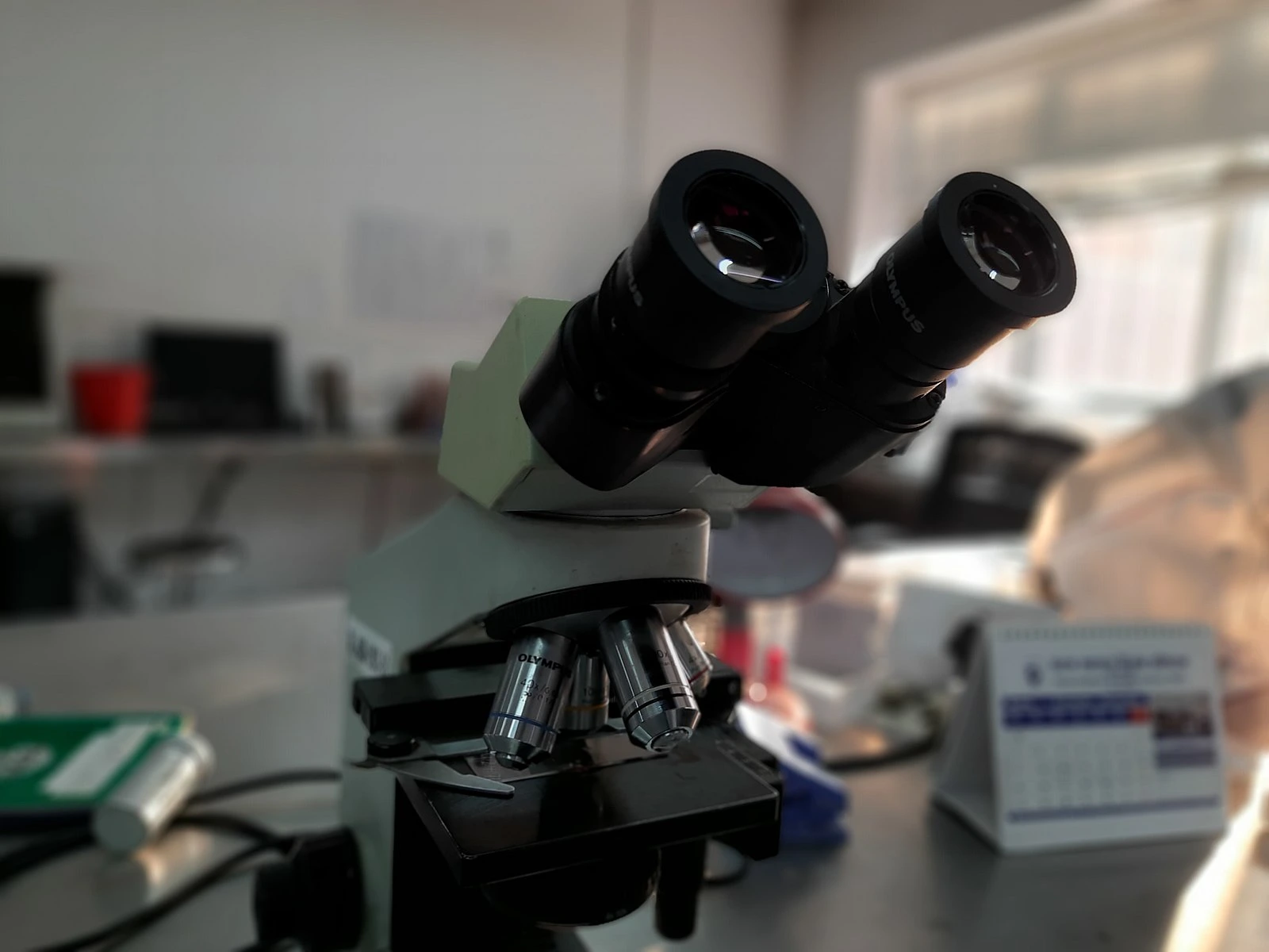
- The image used is licensed under the Creative Commons Attribution-Share Alike 4.0 International license. Author : Ajay Kumar Chaurasiya, Source : Own work (Wikimedia Commons).
*This article is an excerpt from the above mentioned book and Medical Sutras does not make any ownership or affiliation claims.