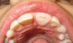
Ellis Class III fracture is confined to enamel and dentin with pulp exposure. The exposed pulp is sensitive to stimuli (eg. air, cold, sweets), while there is normal mobility and no sensitivity to percussion or palpation.
The treatment depends on the size and duration of pulp exposure and the stage of tooth development (open or closed apices).

Direct Pulp Capping
It involves placement of CaOH2 over the exposed pulp, followed by restoration, so that there is no leakage and further trauma to the underlying healthy pulp.
Rationale: Dental pulp may survive and repair small injuries even in the presence of few bacterias, if an adequate seal is provided.
Indications
- If the patient is seen within 24 hours after the injury.
- If the pulp exposure is small (less than 0.5mm).
- If sufficient crown remains to retain a temporary restoration, to support the capping material and prevent the ingress of oral fluids.
Pulpotomy
It involves removal of the damaged and inflammed pulp upto the level of clinically healthy pulp, followed by placement of calcium hydroxide dressing and restoration.
Rationale
- Mild inflammation produces reparative dentin, that forms a bridge between the healthy radicular pulp and the coronal restoration.
- Also, preserving vitality of the radicular pulp ensures normal apexogenesis (root-end development and calcification) in immature teeth.
Indications
- Larger pulp exposure with moderate haemorrhage and healthy radicular pulp.
- The patient is seen within 48 hours.
- Patient did not seek treatment until several hours or days after the injury (even if a small pulp exposure exists).
Shallow/Partial/Cvek's Pulpotomy
- It involves removal of only a portion of the coronal pulp.
- Recommended if the coronal pulpal inflammation is not widespread and if a deeper access opening is not required to help retain the coronal restoration.
Conventional/Cervical/Complete Pulpotomy
- It involves complete removal of the coronal pulp.
- Indicated when the pulpal inflammation extends beyond the pulp chamber.
- In case of trauma to a mature permanent tooth (closed apex) that has caused both a pulp exposure and root fracture.
Pulpectomy
The damaged pulp tissue is removed in entirety to the level of 1-2 mm from the apical foramen.
Indications
- The pulp is degenerated, putrescent or of questionable vitality.
- The duration of exposure is more than 72 hours.
- The final restoration will require the use of post and core for retention.
In case of mature teeth (closed apex), pulpectomy is followed by RCT and restoration, as the closed apex provides a satisfactory situation to achieve an adequate apical seal.
However, in case of immature teeth (open apex), apexification procedure should precede conventional RCT (using GP and sealer), since there is lack of an apical barrier against which the filling can be condensed.
Prognosis
Follow up: Clinical and radiographic evaluations are necessary after 6-8 weeks, 3 months, 6 months and 1 year.
Favourable Outcomes
- Asymptomatic.
- Positive response to pulp sensitivity testing.
- Good quality restoration.
- Continued root development in immature teeth.
Unfavourable Outcomes
- Symptomatic.
- Discolouration.
- Pulp necrosis and infection.
- Apical periodontitis.
- Lack of further root development in immature teeth.
- Loss or breakdown of restoration.
Points to Note
- The tooth should be evaluated for any possible luxation injury or root fracture, especially if tenderness is present.
- The immediate objective is the selection of a procedure that would maintain vitality of the pulp.
- Some experts on pulp therapy recommend conventional pulpectomy and root canal fillings for all teeth treated with CaOH2 pulpotomy, soon after the root apices close. This is to prevent an exaggerated calcific response that may result in total obliteration of the root canal (calcific metamorphosis or calcific degeneration).
- Blunderbuss Canal: Refers to the root canal of an immature tooth in which lumen is largest at the apex and smallest in the cervical area.
References
-
McDonald and Avery's Dentistry for the Child and Adolescent (11th Edition) -Mosby (2021). https://amzn.to/3vZh9m7
-
Grossman's Endodontic Practice (14th Edition) - V. Gopikrishna (editor) - Wolters Kluwer (India) Pvt. Ltd. https://amzn.to/3UrB2MP
-
Bourguignon C, Cohenca N, Lauridsen E, et al. International Association of Dental Traumatology guidelines for the management of traumatic dental injuries: 1. Fractures and luxations. Dent Traumatol. 2020;36:314–330. https://doi.org/10.1111/edt.12578.
-
The image used in cover is licensed under the Creative Commons Attribution-Share Alike 3.0 Unported license.
- Description : A broken upper front tooth showing the pink of the pulp.
- Author : James Heilman, MD.
*This article is an excerpt from the above mentioned books and Medical Sutras does not make any ownership or affiliation claims.