Calcifying Odontogenic Cyst (COC) is a rare developmental odontogenic cyst characterised by ameloblastoma-like lining epithelium and a variable number of ghost cells and spherical calcifications.
Cyst or Tumor ?
- 2005 : WHO classified it as a benign odontogenic tumor, termed as Calcifying Cystic Odontogenic Tumor (CCOT).
- 2017 : WHO reclassified it as cyst, since, clinically most cases behave as non-neoplastic lesions.
Clinical Features
-
Site : Occurs with equal frequency in the maxilla and mandible, most often anterior to first molar.
-
Age : Mostly diagnosed in 20-40 year age group.
-
Intra-osseous COC :
- Presents as slow growing, painless swelling of the jaws.
- In some cases, there may be perforation of cortical plate over the area of expansion, resulting in palpable cystic mass and discharge.
- On aspiration, viscous, granular, yellow fluid is found.
-
Extra-osseous COC : Presents as a localised gingival swelling.
Radiographic Features
- Well-defined, unilocular or multi-locular radiolucency.
- May contain flecks of calcification, seen as radiopaque irregular or tooth-like structures.
- Root resorption or tooth displacement is seen occasionally.
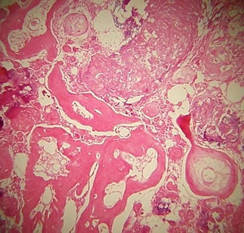
Histologic Features
- The cyst is lined by odontogenic epithelium of varying thickness and is composed of a cuboidal or columnar, palisaded basal cell layer (ameloblastoma-like cells).
- The overlying stellate reticulum-like layer also shows resemblance to that of ameloblastoma.
- Ghost cells : Pale, swollen eosinophilic cells with a hole centrally (loss of nucleus and nuclear membrane).
- The ghost cells often stack up in layers with calcification in a patchy fashion.
- Dentinoid (Dentine-like matrix or mineralised tissue) : Seen in the fibrous cyst wall adjacent to the epithelial lining (Induced where the keratin-like material comes in contact with connective tissue).
Differential Diagnosis
- Fibrous dysplasia (Initial stage) : More common in maxilla and has poorly defined borders that appears as mottled or smoky defined borders on radiograph.
- Odontoma (Partially calcified) : Appears within the capsule.
- Adenomatoid odontogenic tumor (Intermediate stage).
- Ossifying fibroma (Initial stage) : Likely to be situated in more inferior position in the mandible and shows 'Chinese letter' shaped islands of bone or calcification distributed throughout the connective tissue (histologically).
- Odontogenic fibroma : Shows odontogenic tissue like cementum (histologically).
- Cementoblastoma : Well defined lesion, attached to the root of tooth.
Management
- Enucleation and curettage is effective and recommended.
- Prognosis is good after enucleation.
Points to Note
- Other names : Calcifying epithelial odontogenic cyst, Keratinising and calcifying odontogenic cyst, Dentinogenic ghost cell tumor, Calcifying cystic odontogenic tumor, Calcifying ghost cell odontogenic cyst, Gorliin's cyst.
- Occasionally (10% cases), COC is found associated with odontogenic tumors, most commonly odontomas.
- The radiographic appearance is non-diagnostic, unless mineralisation is present.
- Ghost cells are formed due to a peculiar form of abnormal keratinisation that results in clusters of pale swollen, eosinophilic cells, with subsequent degeneration and loss of the nucleus, forming the central hole.
References
-
Contemporary Oral Medicine A Comprehensive Approach to Clinical Practice, Camile S. Farah, Ramesh Balasubramaniam, Michael J. McCullough, Springer.
-
Cawson's Essentials of Oral Pathology and Oral Medicine (9th Edition), E. W. Odell, Elsevier.
-
Textbook of Oral Medicine (3rd Edition), Anil Govindrao Ghom, Savita Anil Ghom (Lodam), Jaypee Brothers Medical Publishers (P) Ltd.
-
Shafer, Hine, Levy Shafer's Textbook of Oral Pathology (7th Edition), Editors - R Rajendran, B Sivapathasundharam, Elsevier.
-
The image used is licensed under the Creative Commons Attribution-Share Alike 3.0 Unported License (CC-BY 2.0).
- Description : Histology of calcified cystic odontogenic tumor.
- Author : Baldanders.
*This article is an excerpt from the above mentioned books and Medical Sutras does not make any ownership or affiliation claims.