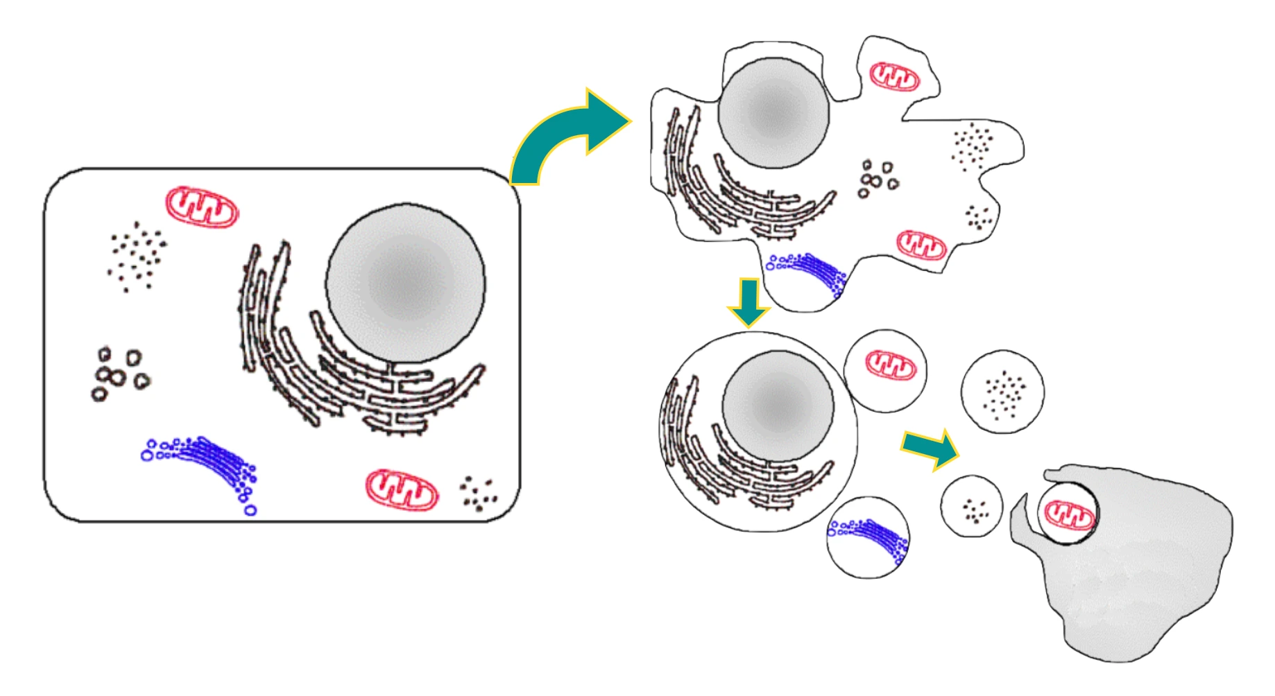
Apoptosis is a pathway of cell death in which cells activate enzymes that degrade their own nuclear DNA and nuclear and cytoplasmic proteins, resulting in break off of the cell fragments (hence, the name apoptosis, meaning falling off).
Etiology
The causes of apoptosis can be grouped as physiologic and pathologic.
Physiologic Apoptosis
There is cell death in normal situations in order to eliminate potentially harmful cells and cells that have outlived their usefulness.
- During embryogenesis and normal development of an organism, presumably due to loss of growth factor signaling.
- Turnover of proliferative tissues e.g., intestinal epithelium, lymphocytes in bone marrow, and thymus (Presumably due to loss of growth factor signaling).
- Involution of hormone-dependent tissues e.g., endometrium (Due to decreased hormone levels, leading to reduced survival signals).
- Removal of excess leukocytes at the end of immune and inflammatory responses (Due to loss of survival signals, as stimulus for leukocyte activation is eliminated).
- Elimination of potentially harmful self-reactive lymphocytes (Induced by both mitochondrial and death receptor pathways).
Apoptosis in Pathologic Conditions
It occurs when cells are damaged beyond repair and needs to be eliminated.
- Severe DNA damage (Activation of proapoptotic proteins by BH3-only sensors).
- Accumulation of misfolded proteins (Activation of proapoptotic proteins by BH3-only sensors, possible direct activation of caspases).
- Infections, exp. certain viral infections (Activation of the mitochondrial pathway by viral proteins and by cytotoxic T lymphocytes, which activate caspases).
Mechanism
In apoptosis, the plasma membrane of the cells remain intact, but it is altered in such a way that the fragments (apoptotic bodies) become highly edible, leading to their rapid consumption by phagocytes.
- It is regulated by biochemical pathways that control the balance between death and survival-inducing signals.
- Involves two pathways: the mitochondrial pathway and the death receptor pathway, that ultimately leads to activation of enzymes called caspases.
- The dead cells and fragments are cleared with little leakage of cellular contents, so apoptosis does not elicit an inflammatory reaction.
Mitochondrial (Intrinsic) Pathway
- Involved in most physiologic and pathologic conditions.
- Mitochondria contain several proteins that are capable of inducing apoptosis, including cytochrome c.
- In healthy cells, the integrity of mitochondrial membranes is maintained by Bcl-2 and related Bcl-xL (produced in response to growth factors and other stimuli). This is done mainly by holding two proapoptotic members of the family i.e., Bax and Bak.
- In case of physiologic apoptosis or pathologic conditions, BH3 proteins are activated, which shift the life-sustaining balance in favor of pro-apoptotic Bak and Bax.
- As a result, Bak and Bax dimerize, insert into the mitochondrial membrane and form channels through which cytochrome c and other mitochondrial proteins leaks out into the cytoplasm.
- The cytochrome c along with other cofactors, activates caspase-9 triggering the caspase cascade and leading to nuclear fragmentation and formation of apoptotic bodies.
Death Receptor (Extrinsic) Pathway
The death receptor pathway is involved in the elimination of self-reactive lymphocytes and in the killing of target cells by some cytotoxic T cells that express FasL.
- Death receptors are surface molecules expressed by several cells. The prototypic death receptors include Type I TNF receptor and Fas (CD95).
- These contain a death domain in their cytoplasmic regions, which mediates interaction with other proteins involved in cell death.
- When the T cells recognise Fas-expressing targets, Fas molecules are crosslinked by Fas ligand (FasL) and bind adaptor proteins via the death domain.
- These then recruit and activate caspase-8 and downstream caspase cascade that cleave numerous targets.
- This ultimately leads to activation of enzymes that degrade the cells' proteins and nucleus.
Clearance of Apoptotic Cells
The dead cells and their fragments are removed by phagocytosis that involves several "eat-me" signals.
- Alteration in plasma membrane: In normal cells, phosphatidylserine is present on the inner leaflet of the plasma membrane, but in apoptotic cells, this phospholipid "flips" to the outer leaflet, where it is recognised by tissue macrophages. This leads to phagocytosis of the apoptotic cells.
- Secretion of proteins: The apoptotic cells secrete soluble factors that recruit phagocytes.
Histopathology
The nuclei of apoptotic cells, in H&E stains, show various stages of chromatin condensation and aggregation and ultimately fragmentation of DNA into nucleosome-sized pieces (karyorrhexis).
- The cells rapidly shrink, form cytoplasmic buds and fragment into apoptotic bodies that are composed of membrane-bound pieces of cytosol and organelles.
- The changes may be histologically undetectable, as these fragments are rapidly extruded and phagocytosed without eliciting an inflammatory response.
References
- Robbins Basic Pathology, 10th edition, Vinay Kumar, Abul K. Abbas, Jon C. Aster, Elsevier.
- The images used is released into the public domain by the copyright holder, under the terms of the GNU Free Documentation License (Source : WIkimedia Commons).
*This article is an excerpt from the above mentioned book and Medical Sutras does not make any ownership or affiliation claims.