The average denture bearing area available for support in edentulous mandible is 14 cm2. Also, mandible is less capable of resisting occlusal forces than maxilla, hence, extra care must be taken to maximise the available contact area in mandible.
Stress Bearing or Supporting areas
The primary stress-bearing area generally have thicker keratinised mucosa and/or underlying cortical bone that lies perpendicular to the occlusal forces and is less susceptible to resorption. It includes,
- Buccal shelf.
The secondary stress-bearing area generally have thin and loose, non-keratinised mucosa, with underlying cancellous bone that lies at an angle to the occlusal forces and is more susceptible to resorption. It includes,
- Retromolar pads
- Residual ridge.
Buccal Shelf
It is the area between the mandibular buccal frenum and the anterior edge of the masseter muscle.
-
Boundaries:
- Medial : Crest of the residual ridge.
- Lateral : External oblique ridge.
- Distal : Retromolar pad.
-
It acts as a suitable primary stress bearing area because,
- The shelf is made up of dense cortical bone.
- It lies at right angle to vertical occlusal forces.
- Muscle attachments on the posterior and lateral borders prevent resorption.
-
With ridge resorption, the denture bearing surface becomes flatter and widens towards the buccal shelf, since, the alveolar ridge of the mandible is significantly mesial to the inferior border of the mandible.
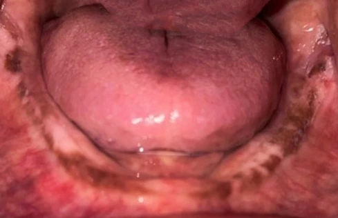
Retromolar Pad
It is a triangular pad of tissue located at distal end of the residual ridge.
- The anterior portion of the triangle is keratinised tissue of the remnant gingiva of the third molar called the pear-shaped pad.
- The posterior aspect of the triangle is composed of thin, nonkeratinised epithelium, loose connective tissue, glandular tissue, fibers of temporalis tendon and the buccinator and superior constrictor muscles, and the pterygomandibular raphe.
- The underlying bone is dense cortical bone because of the muscle attachments and is resistant to resorption.
- The denture should cover the retromolar pad because of the support and lack of long-term cortical bone resorption.
- To avoid displacement of the denture, the muscles should be activated during border molding by opening wide and closing against pressure.
Residual Ridge
- It is considered as secondary stress bearing area.
- The underlying bone is cancellous and covered by fibrous connective tissue, hence not favorable as primary stress bearing area.
Relief Areas
- Secondary stress bearing areas.
- Mandibular tori.
- Retromylohyoid ridge.
- Mental foramen.
- Genial tubercles.
- Undercuts or sharp bony prominence on ridges
Torus Mandibularis
It refers to the bony prominence made of dense cortical bone, usually found on the lingual aspect of mandible near the first and second premolars.
- Lies midway between the soft tissues of the floor of the mouth and crest of the alveolar process.
- May extend posterior to the molar area.
- Small tori may be accommodated by providing relief in the denture. However, they usually need surgical removal for proper extension of the denture in the floor of mouth.
Mylohyoid Ridge
It refers to the bony prominence along the lingual aspect of the mandible, present at the same level as the external oblique ridge.
- Provides attachment the mylohyoid muscle.
- It is most prominent posteriorly near the retromolar pad, that often requires relief in the denture.
- The posterior prominence may require surgical reduction if combined with the posterior torus.
Mental Foramen
It lies below the alveolar ridge, but may be positioned at the crest of the ridge due to continued resorption of the ridge.
- The mental nerve exits the mental foramen and may be compressed by the denture, resulting in pain or altered sensation in the lip.
Genial Tubercles
It is a dense cortical prominence at the inferior border of the mandible at the lingual midline.
- Represents the muscle attachment of the genioglossus muscles.
- Need to be considered in the denture border, when there is extensive resorption of the residual ridge.
Peripheral / Limiting Structures
The peripheral or limiting structures consists of the following :
- Labial border: Labial vestibule.
- Buccal border: Buccal vestibule.
- Distal extension: Ramus and pterygomandibular raphe.
- Lingual border: Mylohyoid muscle.
- Posterolingual border: Retromylohyoid fossa/curtain.
Labial Vestibule
- Extends from the labial frenum to the buccal frenum.
- The mentalis muscle inserts very close to the crest of the ridge in this area and limits the border extension in length and width.
- The labial frenum contains fibers of the orbicularis oris muscle.
- Both orbicularis oris and mentalis muscles are very active and opening wide will thin the dimensions of the denture border.
Buccal Vestibule
- Extends posteriorly from the buccal frenum to the posterior lateral aspect of the retromolar pad.
- The denture is very wide in this area.
- The mucolabial fold consists of the buccinator muscle, that runs horizontal to the denture and depending on the vertical position of the mucobuccal fold, the denture may cover the entire external oblique ridge.
- Vertical fibers of the masseter muscle are present along the distobuccal border of the buccal vestibule.These fibers are activated by occlusal forces and cause a bulge in the buccinator muscle, creating the masseteric notch in the postero-lateral denture border.
- During border molding, masseter muscle can be activated by pushing downward on the patient's chin while the patient attempts to close the mouth against this pressure.
Ramus and Pterygomandibular Raphe
- Limits the distal extension of the denture, as the sharpness of the internal oblique ridge (mylohyoid ridge) and the external oblique ridge meet at the ascending ramus.
- If the denture border contacts the ascending ramus, it may traumatically trap tissues against the ramus.
- Pterygomandibular raphe is composed of fibers of buccinator and superior constrictor muscles.
Lingual Border/Mylohyoid Muscle
- Defined by the mylohyoid muscle along the entire length of the mandible.
- Posteriorly, resorption of the alveolar ridge effectively brings the internal oblique ridge and its muscle attachments to a position nearer the residual ridge crest.
- Along the lingual border the mylohyoid muscle is at right angle to the border, and it therefore flattens the border with its muscle action and also can slightly pull the border away from the boney lateral wall of the mandible.
- In the anterior, the muscle meets in the midline in a fan shape.
- In the anterior the sublingual gland overlies the mylohyoid muscle.
- The denture border can be quite wide in the midbody where the mylohyoid begins to attach closer to the ridge crest.
- The value of the border's maximum lingual border extension is lateral stability of the denture and reduction of food collection under the denture.
Retromylohyoid Fossa & Retromylohyoid Curtain
- Retromylohyoid fossa presents as an undercut in the postero-inferior part of the mylohyoid ridge.
- The mylohyoid muscle is not active in this area and the denture flange turns laterally into the retromylohyoid fossa, resulting in S shape of the lingual denture border.
- Retromylohyoid curtain forms the posterior border of retromylohyoid fossa and consists of mucosa covering the palatoglossus muscle (posteromedially) and the superior constrictor muscle (posterolaterally).
- The medial pterygoid muscle lies behind the superior constrictor muscle, and contraction of the medial pterygoid muscle can cause a bulge in the wall of the retromylohyoid curtain.
- Protruding the tongue also elevates the curtain in this area and will unseat the denture.
Points to Note
- Resorption of the mandibular residual ridge results in buccal positioned, flatter and wider denture-bearing area, since the mandibular dentition is positioned significantly lingual to the basal bone of the mandible.
- The buccinator muscle extends from the modiolus anteriorly, and its fibers end in the pterygomandibular raphe and are attached laterally along the external oblique ridge.
- The vertical fibers of the masseter muscle, originates on the zygomatic arch and attach to the mandible lateral to the buccinator fibers.
- Swallowing moves the superior constrictor and palatoglossus muscle forward and denture patients may complain of pain on swallowing.
- In a denture retained with implants, overextension in the retromylohyoid fossa or along the mylohyoid muscle may cause an ulcer because the denture cannot unseat.
References
- Prosthodontic Treatment for Edentulous Patients Complete Dentures and Implant-supported Prostheses (13th edition) , Zarb, Hobkirk, Eckert, Jacob, Mosby Elsevier.
*This article is an excerpt from the above mentioned book and Medical Sutras does not make any ownership and affiliation claims.