Amyloidosis is a condition in which there is extracellular deposition of fibrillar proteins in body organs, that leads to abnormal protein build-up in the tissues, resulting in tissue damage and organ dysfunction.
The deposits were referred as amyloid since they bind a wide variety of proteoglycans and glycosaminoglycans, which contain charged sugar groups and give the deposits staining characteristics resembling those of starch (amylose).
Composition & Forms of Amyloid
All amyloid deposits are composed of non-branching fibrils (95% consists of fibril proteins, 5% glycoproteins), that are formed of intertwined polypeptides in a beta pleated sheet conformation. The three most common form of amyloid are:
-
AL (Amyloid light chain) amyloid
- Made up of complete immunoglobulin light chains, the amino-terminal fragments of light chains or both.
- AL protein does not loose affinity for Congo red after incubation of tissue sections with KMnO4.
-
AA (Amyloid-associated) amyloid
- Composed of non-immunoglobulin protein (8500-dalton protein).
- Derived by proteolysis from a larger precursor in the blood, called Serum Amyloid Associated (SAA) protein (synthesised in the liver).
- AA protein loses affinity for Congo red after incubation of tissue sections with KMnO4.
-
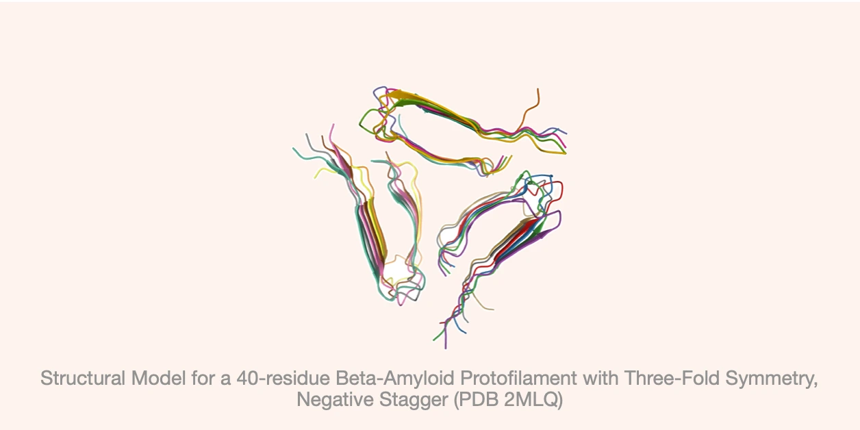
Beta-amyloid protein : Composed of 4000-dalton peptide, that is derived by proteolysis from a much larger transmembrane glycoprotein, called amyloid precursor protein.
Mechanism of formation
Amyloidosis results from abnormal folding of proteins, which assume a beta pleated sheet conformation, aggregate, and deposit as fibrils in extracellular tissues.
-
Normally, the intracellular misfolded proteins are degraded in proteasomes and extracellular protein aggregates are taken up and degraded by macrophages.
-
However, in amyloidosis, these quality control mechanisms fail and fibrillar proteins accumulate outside of cells.
-
The proteins that form amyloid fall into two categories:
- Normal proteins that have an inherent tendency to associate and form fibrils, particularly when produced in increased amounts
- Mutant proteins that are prone to misfolding and aggregation.
Classification
-
Systemic (Generalised), involving several organ systems.
- Primary amyloidosis (Type B), when it is associated with a clonal plasma cell proliferation.
- Secondary amyloidosis (Type A), occur as a complication of an underlying chronic inflammatory, genetic diseases, syndromes such as familial mediterranean fever.
-
Localised to a single organ, such as the heart.
-
Hereditary or familial amyloidosis : Constitutes a separate, heterogeneous group with several distinctive patterns of organ involvement.
-
Type C amyloid : Includes amyloid of aging, localised non-specific amyloid, amyloid adjacent to APUD (Amine precursor uptake and decarboxylase) i.e, pheochromocytoma.
Primary Systemic Amyloidosis
- This is the most common form of amyloidosis, composed of AL type amyloid and usually systemic in distribution.
- Caused by a clonal proliferation of plasma cells that synthesise abnormal Ig molecules.
- It is referred as primary, since, the clinical features are derived solely from the effects of amyloid deposition rather than formation of tumor masses.
- In virtually all cases, monoclonal immunoglobulins or free light chains or both can be found in the blood or urine.
- Most patients with AL amyloid do not have multiple myeloma or any other overt B cell neoplasm.
- Occurs in 5-15% of individuals with multiple myeloma (a plasma cell tumor characterised by excessive production of free immunoglobulin light chains).
Reactive Systemic Amyloidosis
- It is composed of AA proteins and occurs secondary to an associated inflammatory condition such as Rheumatoid arthritis (3% cases), Ankylosing spondylitis, and Inflammatory Bowel Disease ( particularly Crohn disease and Ulcerative colitis).
- It may also occur in association with certain cancers, the most common being Renal cell carcinoma and Hodgkin lymphoma.
- Heroin abusers who inject the drug subcutaneously also have a high occurrence rate of generalised AA amyloidosis. The chronic skin infections, which cause the "skin-popping" associated with injection of narcotics, seem to be responsible for the amyloidosis.
- In AA amyloidosis, there is sustained elevation of SAA levels due to long standing inflammation, since, Serum Amyloid Associated protein synthesis by liver cells is stimulated by cytokines such as IL-6 and IL-1 that are produced during inflammation.
Localised Amyloidosis
-
The amyloid deposits are limited to a single organ or tissue without involvement of any other site in the body.
-
It may produce grossly detectable nodular masses or may be evident only on microscopic examination.
-
Nodular deposits of amyloid are often encountered in the lung, larynx, skin, urinary bladder, tongue, and the region about the eye.
-
Endocrine amyloid
- Composed of microscopic deposits of localised amyloid, found in certain endocrine tumors, such as medullary carcinoma of the thyroid gland, Islet tumors of the pancreas, pheochromocytomas and undifferentiated carcinomas of the stomach, and in the Islets of Langerhans in individual with type 2 DM.
- The amyloid proteins seem to be derived either from polypeptide hormones (e.g., medullary carcinoma) or from unique proteins (e.g., islet amyloid polypeptide).
Heredofamilial Amyloidosis
-
Rare and occurs in limited geographical areas.
-
Most commonly found in Familial Mediterranean fever (autosomal recessive condition),
- AA type amyloid is seen, suggesting that it is related to the recurrent bouts of inflammation.
- auto-inflammatory syndrome associated with excessive production of the cytokine IL-1 in response to inflammatory stimuli
- characterised by attacks of fever accompanied by inflammation of serosal surfaces that manifests as peritonitis, pleuritis and synovitis.
-
Also found in autosomal dominant familial disorders,
- characterised by deposition of amyloid made up of fibrils derived from mutant transthyretin (TTR).
- Specific TTR mutant polypeptides tend to form amyloid in different peripheral organs:
- Familial amyloidotic polyneuropathies : Deposits are seen mainly in peripheral nerves.
- Cardiac amyloidosis : Cardiac deposits predominate.
Macroscopic findings / Morphology
- Amyloid may be appreciated macroscopically when it accumulates in large amounts.
- The organ is frequently enlarged and the tissue appears gray and has a waxy, firm consistency.
- Most commonly affected organs : Kidneys, Heart, GIT, Liver, Spleen. Tongue (macroglossia) and gingiva.
- Gingival biopsy may be used for diagnosis.
- Cut surface painted with Iodine and sulfuric acid: Mahogany Brown staining.
Histological Features
-
Histologically, the amyloid deposition is always extracellular and begins between cells, often closely adjacent to basement membrane. As the amyloid accumulates, it encroaches on the cells, surrounds and destroys them.
-
In primary amyloidosis (associated with plasma cell proliferation), perivascular and vascular deposits are common.
-
Light microscopy (H & E stain): Amyloid appears as an amorphous, eosinophilic, hyaline, extracellular substance.
-
To differentiate amyloid from other hyaline materials (e.g., collagen, fibrin), Congo red can be used:
- Gives a pink or red color to tissue deposits under ordinary light.
- Polarised light microscopy : Green birefringence of the stained amyloid, due to the crossed beta-pleated sheet configuration of amyloid fibrils
-
Electron microscopy (confirmatory): Seen as amorphous non-oriented thin fibrils.
-
Mass spectroscopy: Most reliable for sub-typing of amyloid.
Points to Note
-
Most common diseases predisposing to amyloidosis:
- Rheumatoid arthritis.
- Myeloma.
- Chronic infections: TB and osteomyelitis, Inflammatory bowel disease (largely eliminated by modern medicine and surgery).
-
Multiple myeloma:
- A plasma cell tumor characterised by excessive production of free immunoglobulin light chains.
- The free, unpaired kappa and lambda light chains (referred to as Bence Jones protein) are prone to aggregation and deposition in tissues as amyloid.
- Not all free light chains are equally likely to produce amyloid, majority of myeloma patients do not develop amyloidosis.
- Lambda light chains are approximately six times more likely to deposit as amyloid than kappa light chains.
-
Hemodialysis-Associated Amyloidosis:
- Seen in patients on long-term hemodialysis where beta-2-microglobulin form amyloid deposits, as it cannot be filtered through dialysis membranes and retained in the circulation.
- However, with new dialysis filters, there is substantial decrease in the incidence.
- Classic features include triad of scapulohumeral periarthritis, carpal tunnel syndrome, and flexor tenosynovitis of the hand.
-
Senile systemic amyloidosis (Amyloid of aging):
- Refers to systemic deposition of amyloid in elderly patients (usually in their 70s and 80s).
- The amyloid is derived from normal TTR.
- The heart is predominantly involved with related dysfunction (hence,previously called senile cardiac amyloidosis).
References
- Robbins Basic Pathology (10th Edition) - Vinay Kumar, Abul K. Abbas, Jon C. Aster, Elsevier.
- Shafer, Hine, Levy Shafer's Textbook of Oral Pathology (7th Edition), Editors - R Rajendran, B Sivapathasundharam, Elsevier.
- The image used is licensed under the Creative Commons Attribution-Share Alike 4.0 International license. Source: Wikimedia Commons, Author : Pierre Hugots. (https://commons.wikimedia.org/wiki/File:Protofilament_of_Beta_Amyloid.jpg)
*This article is an excerpt from the above mentioned books and Medical Sutras does not make any ownership or affiliation claims.