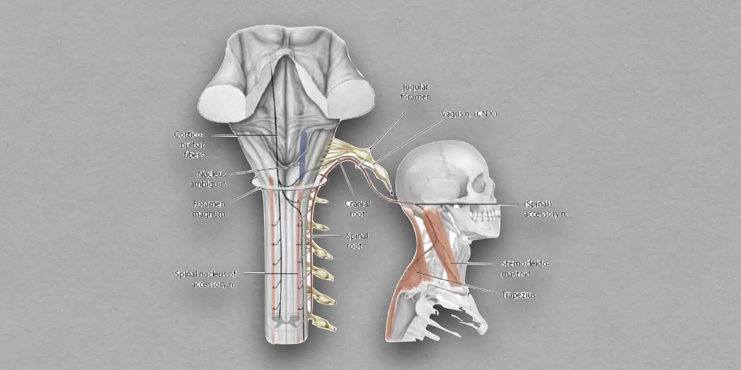
It is the 11th cranial nerve that is purely motor and consists of two roots: cranial and spinal.
- Cranial root: Accessory to the vagus and its fibres are distributed through the vagus nerve.
- Spinal root: Have an independent course and is referred as spinal accessory nerve.
Functional Components
Special Visceral Efferent Fibres
- Arises from the nucleus ambiguus and form the cranial root.
- Provides motor supply to the muscles of soft palate, pharynx, and larynx.
General Somatic Efferent Fibres
- Arises from the spinal nucleus of accessory nerve, in the ventral horns of the upper five spinal segments,
- Forms the spinal root.
- Provides motor supply to the sternocleidomastoid and trapezius muscles.
Anatomy & Pathway
Origin
- Cranial root arises by 4-5 rootlets from the posterolateral sulcus of the medulla between the olive and inferior cerebellar peduncle.
- Spinal root arises by a number of rootlets from the lateral aspect of the spinal cord (upper five cervical spinal segments).
Intracranial Course
- The rootlets of cranial root are attached in line with the rootlets of the vagus nerve above. They unite together to form a single trunk that runs laterally along with the glossopharyngeal and vagus nerve, and, reach the jugular foramen.
- The rootlets of spinal root unite to form a single trunk that ascends in the vertebral canal to enter the cranial cavity through the foramen magnum behind the vertebral artery.
- In the jugular foramen, both the cranial and spinal root fuse and forms a combined trunk. The trunk leaves the cranial cavity through the middle compartment of the jugular foramen enclosed in the dural sheath, along with the vagus nerve.
Extracranial Course
The cranial and spinal roots separate immediately after exiting the cranial cavity.
- Cranial root joins the vagus nerve below its inferior ganglion and is distributed through the branches of the vagus nerve.
- Spinal root descends vertically downward between the internal jugular vein and internal carotid artery.
- It turns downward and backward across the internal jugular vein toward the carotid triangle, at a point midway between the angle of mandible and the mastoid process.
- Then, it crosses in front of the transverse process of the atlas under the posterior belly of digastric and occipital artery.
- The spinal nerve pierces the sternocleidomastoid muscle at the junction of its upper 1/4th with lower 3/4th. It passes through the muscle and emerges through its posterior border.
- It then enters the posterior triangle, runs downward and backward underneath the fascial roof of the posterior triangle, parallel to the fibres of levator scapulae.
- It leaves the posterior triangle by passing deep to the anterior border of trapezius about 5 cm above the clavicle.

Distribution
Cranial Root
- All muscles of the palate except the tensor palati and tensor tympani (supplied by the mandibular nerve).
- All muscles of the pharynx except the stylopharyngeus (supplied by the glossopharyngeal).
- All intrinsic muscles of larynx.
Spinal Root
- Sternocleidomastoid muscle along with C2 and C3 spinal nerves.
- Trapezius muscle along with C3 and C4 spinal nerves.
Clinical Significance
The spinal accessory nerve can get damaged in case of:
- Fracture of the base of the skull through jugular foramen.
- Stab wounds in the neck.
- Surgical removal of cervical lymph nodes.
Unilateral lesion of spinal accessory nerve proximal to sternocleidomastoid causes:
- Ipsilateral paralysis of sternocleidomastoid: Inability to tilt the head toward the ipsilateral shoulder and turn face toward the opposite side.
- Paralysis of trapezius: Inability to shrug shoulder against resistance.
Spasmodic torticollis
- Characterised by clonic spasms of sternocleidomastoid.
- May result from irritative central lesions of spinal accessory nerve.
Clinical Testing of Spinal Accessory Nerve
- Sternocleidomastoid: Ask the patient to turn his/her face to the opposite against the resistance offered by the examiner's hand. In normal case, the patient can do it and sternocleidomastoid stands out prominently.
- Trapezius: Ask the patient to shrug his/her shoulder against resistance.
References
| [](https://amzn.to/3Ixnict)
Textbook of Anatomy Head, Neck, and Brain (Volume III), Vishram Singh |
- Image Credit: Diyorbek 6504 (Source: Wikimedia Commons, Creative Commons Attribution-Share Alike 4.0 International license).
*This article is an excerpt from the above mentioned sources and Medical Sutras does not make any ownership or affiliation claims.