It is the sixth cranial nerve, that is purely motor and supplies only one muscle i.e., lateral rectus muscle.
- It abducts the eye, hence, the name abducent.
- Also called lover's nerve, since, the lateral rectus muscle was used for non-verbal communication between lovers in older times.
- Longest and thinnest nerve.
- Most susceptible to damage of all cranial nerves due to increased intracranial pressure.
Functional Components
General Somatic Afferent Fibres
- Carry proprioceptive sensations from lateral rectus.
- Terminate in the mesencephalic nucleus of the trigeminal nerve.
General Somatic Efferent Fibres
- Originate from the abducent nucleus in the pons.
- Supply the lateral rectus muscle.
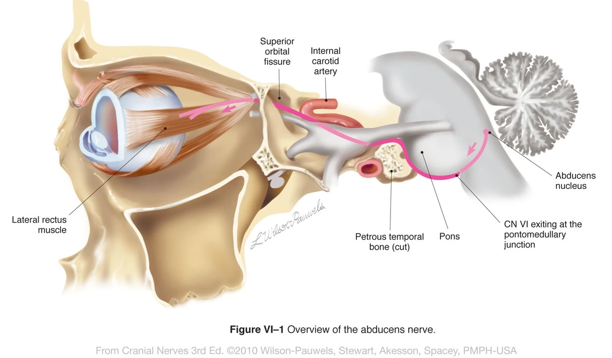
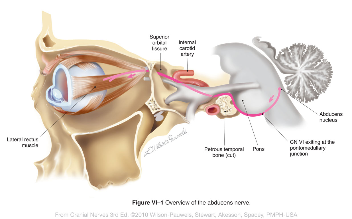
Anatomy & Pathway
- Arises at the lower border of the pons opposite the pyramid of medulla.
- Runs upward, forward and laterally dorsal to the anterior cerebellar artery and pierces the dura mater over the clivus, inferolateral to the dorsum sellae.
- Then, it passes through the medial wall of the inferior petrosal sinus and arches forward directly over the ridge of petrous temporal bone, under the petroclinoid ligament.
- It enters the fibro-osseous canal (Dorello's canal) formed by the apex of the petrous temporal bone and petroclinoid ligament (Gruber's ligament).
- The nerve then enters the cavernous sinus by piercing the posterior wall close to the floor of the sinus.
- In the cavernous sinus, it runs forward inferolateral to the internal carotid artery and enters the orbit through the superior orbital fissure within the tendinous ring lateral to two divisions of oculomotor and nasociliary nerves.
- In the orbit, it runs forward, toward the lateral side to enter the orbital surface of the lateral rectus muscle which it supplies.
Clinical Significance
The abducent nerve is stretched and damaged during increased intracranial pressure due to the descent of brainstem. Consequently, the nerve is cut by the sharp edge of the petrous temporal bone.
This leads to the paralysis of lateral rectus muscle and the following clinical findings:
- Convergent squint: Due to unopposed action of medial rectus.
- Inability to abduct the eye.
- Diplopia: Double vision with maximum separation of two images while looking toward the paralysed side.
References
| [](https://amzn.to/3Ixnict)
Textbook of Anatomy Head, Neck, and Brain (Volume III), Vishram Singh |
*This article is an excerpt from the above mentioned sources and Medical Sutras does not make any ownership or affiliation claims.