Epithelial tissues form protective barriers and specialized secretory units, classified by the number of cell layers (simple or stratified) and cell morphology (squamous, cuboidal, or columnar). The oral mucosa is a highly specialized stratified squamous epithelium that undergoes precise maturation processes—such as keratinization—to resist mechanical and chemical stress, relying entirely on robust cellular junctions and a specialized basement membrane for structural integrity and connective tissue anchorage.
🔍 Detailed Breakdown
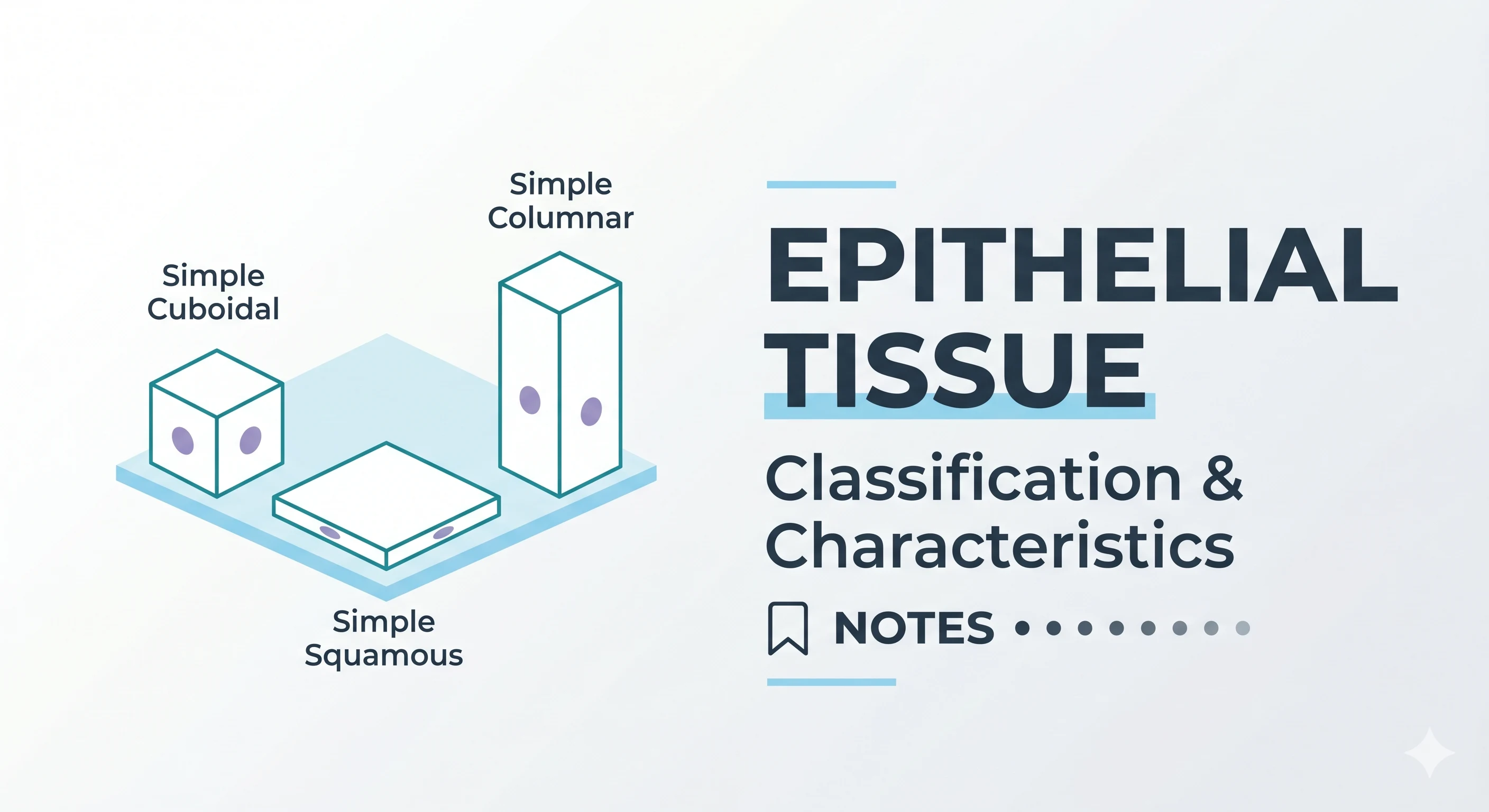
- Epithelial Classification:
- Simple epithelia consist of a single layer of cells, typically cuboidal or columnar, facilitating secretion and absorption (e.g., salivary gland acini and ducts, or differentiating ameloblasts).
- Stratified epithelia feature multiple layers designed for protection. The oral cavity is lined by stratified squamous epithelium, where the deepest progenitor cells are cuboidal or columnar, but the surface cells mature into flattened squamous (scale-like) profiles.
- The Concept of Keratinization: A directed maturation process of keratinocytes. As cells migrate from the basal layer to the surface, they undergo terminal differentiation:
- Stratum Basale: A single layer of highly mitotic cuboidal/columnar cells.
- Stratum Spinosum (Prickle Cell Layer): Larger polyhedral cells synthesizing abundant cytokeratins (intermediate filaments).
- Stratum Granulosum: Flatter cells containing basophilic keratohyalin granules. These granules release filaggrin, a protein that aggregates and cross-links the cytokeratin filaments.
- Stratum Corneum (Keratinized Layer): The surface layer consisting of dehydrated, tightly packed squames. In orthokeratinization, all organelles and nuclei are lost. In parakeratinization (common in the gingiva), cells retain shrunken, pyknotic nuclei.
- The Basement Membrane (Basal Lamina): A 50 to 100 nm thick extracellular matrix interface separating the avascular epithelium from the connective tissue (lamina propria).
- Lamina Lucida: An electron-clear zone directly beneath the basal cells containing laminin, integrins, and bullous pemphigoid antigen.
- Lamina Densa: A dark, electron-dense zone composed of a "chicken-wire" network of Type IV collagen, perlecan (heparan sulfate proteoglycan), and nidogen.
- Lamina Fibroreticularis: Features Type VII collagen forming anchoring fibrils that loop into the lamina densa to firmly tether the epithelium to the underlying connective tissue.
- Cellular Junctions (The Mechanical Linkage):
- Desmosomes (Macula Adherens): Cell-to-cell junctions providing tensile strength. They utilize transmembrane cadherins (desmoglein and desmocollin) linked to dense intracellular attachment plaques (containing desmoplakin and plakoglobin). Intermediate filaments (tonofilaments/cytokeratins) insert directly into these plaques.
- Hemidesmosomes: Cell-to-matrix junctions anchoring basal cells to the lamina lucida. They utilize transmembrane integrin α6β4 and collagen XVII (BP180), which connect to extracellular laminin-332 and intracellular plaque proteins (BP230 and plectin), anchoring the cellular intermediate filaments to the basement membrane.
🩺 Clinical Relevance
- Clinical Highlight: The structural integrity of the oral mucosa relies entirely on the precise molecular assembly of its cellular junctions and basement membrane. Autoantibodies attacking desmosomal cadherins (such as desmoglein-3) destroy cell-to-cell adhesion, causing intraepithelial splitting and the severe blistering disease pemphigus vulgaris. Conversely, autoantibodies targeting hemidesmosomal proteins (collagen XVII/BP180) cause bullous pemphigoid, resulting in the detachment of the entire epithelium from the underlying connective tissue at the lamina lucida.
📝 Exam High-Yield Points
- Cytokeratins (7–11 nm diameter) are the defining intermediate filaments of all epithelial cells (keratinocytes), distinguishing them from mesenchymal cells.
- Desmosomes connect epithelial cells to one another, while hemidesmosomes strictly anchor basal epithelial cells to the underlying basement membrane. Both junction types securely anchor to intracellular intermediate filaments (tonofilaments).
- The lamina densa is built on a primary scaffold of Type IV collagen, while Type VII collagen forms the anchoring fibrils that bind the basal lamina to the lamina propria.
- The "spiny" or "prickle" appearance of the stratum spinosum is an artifact of histologic processing, where cells shrink away from one another but remain firmly tethered at their desmosomal junctions.
References:
- Nanci, A. Ten Cate’s Oral Histology: Development, Structure, and Function. Elsevier (2018).
- Kumar, G. S. Orban's Oral Histology & Embryology. Elsevier (2019).