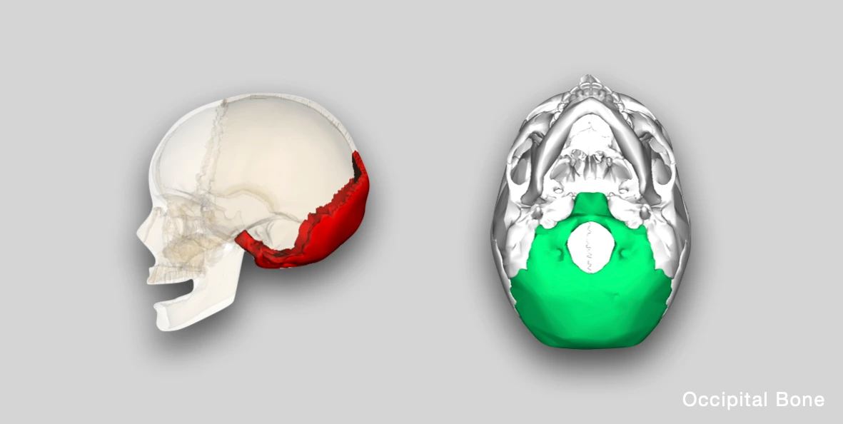
The occipital bone forms the posterior and inferior part of the skull. It consists of four parts i.e., one squamous, two condylar/lateral and one basilar part, arranged around a large opening called foramen magnum.
While the surfaces can be studied separately for each part, the borders and angles are presented together.
Squamous Part
The squamous part is an expanded plate above and behind the foramen magnum.
It comprises of two surfaces (external and internal), four borders (two superior and two inferior), and three angles (one superior and two lateral).
External Surface
It is convex and presents the following features:
- External occipital protuberance: It is a bony prominence along the midline, situated halfway between lambda and foramen magnum.It marks the junction of head and neck, and provides attachment to nuchal ligament and trapezius muscle.
- Inion: It refers to the most prominent point on the external occipital protuberance.
- Occipital point: It is a median point that is farthest from the glabella, and is located a little above the inion.
- External occipital crest: It is a ridge along the midline descending from the external occipital protuberance to the foramen magnum. It provides attachment to nuchal ligament, and also referred as median nuchal line.
- Superior nuchal lines: These are curved bony ridges extending laterally from the occipital protuberance.
- Inferior nuchal lines: These are curved bony ridges passing laterally from the middle of external occipital crest.
- Highest/Supreme nuchal lines: These are faint bony ridges seen in some cases, about 1 cm above the superior nuchal lines.
- Interparietal bone (Inca bone): It is a large triangular bone occasionally present at the apex of the squamous part. It represents the membranous part of the occipital bone that has failed to fuse with the rest of the bone.
 Source: Henry Gray (1918) Anatomy of the Human Body
Internal Surface
It is concave and shows the following features:
-
Internal occipital protuberance: It is a bony prominence located near the center of the squamous part, directly opposite to the external occipital protuberance. It is the point of intersection of the four divisions of cruciform eminence, and is associated with the confluence of dural venous sinuses.
-
Internal occipital crest: It is a bony ridge along the midline that runs downward from the internal occipital protuberance to the posterior margin of foramen magnum. It bifurcates near the foramen magnum and gives attachment to the falx cerebelli.
-
Vermian fossa: It is a shallow midline depression at the inferior end of internal occipital crest, near the posterior edge of foramen magnum. It lodges the inferior part of cerebellar vermis.
-
Grooves of dural venous sinuses: These radiate from the internal occipital protuberance and include:
- Sagittal sulcus/groove: It lodges the superior sagittal sinus and extends upward towards the superior angle.
- Transverse sulcus/groove: It is the groove for transverse sinus that runs laterally, one on each side.
- Internal occipital crest: It houses the occipital sinus and extends inferiorly towards the foramen magnum.
-
Cruciform/Cruciate eminence: It refers to the cross-shaped arrangement of the four grooves of the dural venous sinuses. It divides the internal surface into four fossae:
- Cerebral fossae (upper two): Lodges the occipital lobes.
- Cerebellar fossae (lower two): Accommodates the hemispheres of the cerebellum.
Source: Henry Gray (1918) Anatomy of the Human Body
Condylar/Lateral Part
The lateral parts of occipital bone are located on either side of the foramen magnum and presents the occipital condyles.
It comprises of two surfaces (superior and inferior), and part of inferior border which articulates with the petrous part of temporal bone.
Superior Surface
- Jugular tubercles: These are two small bony protuberances seen at the junction of condylar and basilar parts, just medial to the jugular foramen. The tubercles arise from the concave inferolateral margins of the clivus and project posterosuperiorly over the hypoglossal canals
- Hypoglossal canal (Anterior condylar canal): It pierces the bone posteroanterior to the jugular tubercle, and runs obliquely forwards and laterally along the line of fusion of basilar and condylar parts.
- Condylar canal (Posterior condylar canal): It opens in the lower part of sigmoid sulcus, and transmits an emissary vein connecting the sigmoid sinus with suboccipital venous plexus.
Inferior Surface
- Occipital condyles: These are kidney-shaped bony prominences that connects the skull and vertebral column. They articulate with the first cervical vertebrae (atlas), forming the atlanto-occipital joint.
- Jugular process: It is a quadrilateral plate of bone that projects laterally from the posterior half of the occipital condyle. Its inferior surface is rough and provides attachment surface to muscles, while the superior surface bears a deep groove which is continuous with the jugular notch.
- Outer Opening of Hypoglossal canal: It lies lateral to the anterior part of occipital condyle and transmits the hypoglossal nerve.
- Condylar fossa: It is a depression just behind the occipital condyle.
- Jugular notch: It is present on the anterior margin of jugular process. It forms the jugular foramen with similar notch present on the petrous temporal bone.
*The jugular foramen transmits the following structures:
- Anterior part: Inferior petrosal sinus and meningeal branch of ascending pharyngeal artery.
- Middle part: 9th, 10th and 11th cranial nerves.
- Posterior part: Internal jugular vein and meningeal branch of occipital artery.
Basilar Part
The basilar part or basiocciput is a wide, quadrilateral-shaped bar of bone, that lies anterior to foramen magnum, and adjacent to the petrous temporal bone.
It comprises two surfaces (superior and inferior) and four borders (one anterior, one posterior, and two lateral).
Superior Surface
- Clivus: It is a shallow gutter (sloping surface) formed by the fusion of basiocciput and the body of sphenoid (incl. dorsum sellae). It slopes downwards and backwards from dorsum sellae to the foramen magnum.
- Petro-occipital fissure: It is a groove present on either side, that separates the clivus from petrous part of temporal bone. It is grooved by the inferior petrosal sinus, and is continuous posteriorly with the jugular foramen.
Inferior Surface
- Pharyngeal tubercle: It is a bony bump, found in the median plane, about 1 cm in front of the foramen magnum. It acts as the attachment point for the pharyngeal raphe, to which all the pharyngeal constrictor muscles are connected.
Borders
There are four borders - two superior and two inferior.
Superior Border
- It extends laterally on both sides from the lambda or superior angle to the lateral angle.
- It articulates with the parietal bone along the lambdoid suture, hence, also referred as lambdoid border.
Inferior Border
- It is present on both sides from the lateral angles to the inferior angle.
- In the squamous part (upper half) , it articulates with the mastoid portion of temporal bone, forming the occipitomastoid suture. Here, it is also referred as the mastoid border.
- In the lower half (condylar and basilar part), it articulates with the petrous part of temporal bone, along the petro-occipital suture.
Angles
There are four angles - one superior, two lateral, and one inferior.
Superior Angle
- It is present at the top of the squamous part where both the superior borders meet.
- It is the meeting point of lambdoid and sagittal sutures (lambda).
Lateral Angles
- These are present on either side at the extremities of the groove for transverse sinus, and forms the meeting point of superior and inferior borders.
- It is also known as asterion (meeting point of lambdoid, occipitomastoid, and parietomastoid sutures).
Inferior Angle
- It is fused to the body of the sphenoid bone, forming the basisphenoid joint.
- The basisphenoid joint is responsible for the growth of skull in length.
Foramen Magnum
It is the largest foramen of skull that is oval in shape and connects posterior cranial fossa (upwards) with the vertebral canal (downwards).
The structures passing through foramen magnum include:
Anterior/Osseoligamentous Compartment
- Apical ligament of dens.
- Vertical band of cruciate ligament.
- Tectorial membrane (Membrana tectoria).
Posterior/Neurovascular Compartment
- Medulla oblongata (lowest part).
- Spinal branch of accessory nerve.
- Vertebral arteries.
- Sympathetic plexus around the vertebral arteries.
- Anterior and posterior spinal arteries.
*The three meninges pass through the posterior part and the above nerve and vessels pass through the subarachnoid space (between arachnoid and pia mater).
Anatomical Position
In the anatomical position, the occipital bone should be held with:
- the concave surface/fossae forwards,
- the condylar processes downwards, and,
- the basilar part anteriorly, with the inferior angle pointing upwards.
 Source: en:Anatomography (Wikimedia Commons)
Applied Anatomy
- The basisphenoid joint is important in the assessment of age of an individual. It is a primary cartilaginous joint that is completely replaced by bone by the age of 25 years.
References
- BD Chaurasia's Human Anatomy Volume 3 (Head & Neck), Eighth edition, CBS Publishers and Distributors Pvt Ltd
- Textbook of Anatomy Head, Neck, and Brain (Volume III), Vishram Singh.
- Radiology of the Jugular Tubercles https://www.ajronline.org/doi/pdf/10.2214/ajr.131.6.1037.
- Cover Image Credit: Source - Anatomography[3] website maintained by Life Science Databases(LSDB). This file is made available under the Creative Commons Attribution-Share Alike 2.1 Japan license.
*This article is an excerpt from the above mentioned sources and Medical Sutras does not make any ownership or affiliation claims.