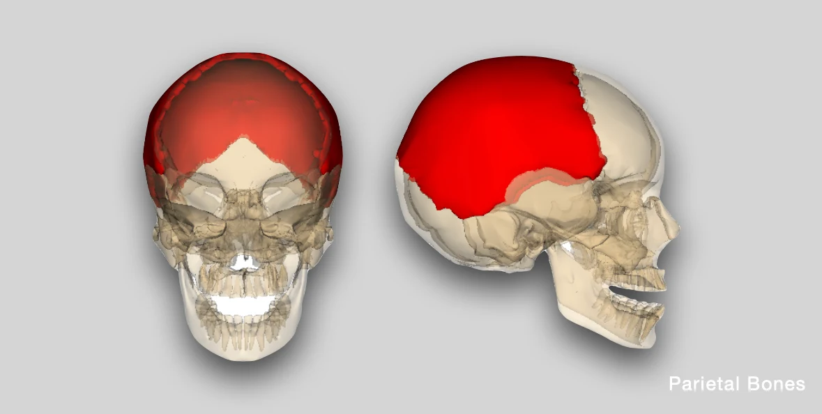
The parietal bone is a flat, quadrilateral, curved bone that forms the roof and sides of the vault of skull.
Surfaces
Outer Surface
The outer surface is smooth and convex, and presents the following features:
- Parietal tuber/eminence: It refers to the area of maximum convexity of the parietal bone.
- Parietal foramen: It lies near the posterior end of sagittal suture, 2.5-4 cm in front of lambda.
- Temporal lines: The inferior temporal line gives origin to the temporalis muscle, and the superior temporal line provides origin to the temporal fascia.

Source: Henry Gray (1918) Anatomy of the Human Body
Inner Surface
The inner surface is concave and presents the following features:
- Sagittal sulcus: It is a shallow depression/groove found along the superior border, and houses/lodges the superior sagittal sinus.
- Granular foveolae (Arachnoid foveolae): These are bony depressions or pits found near the sagittal sulcus, along the length of the superior sagittal sinus. These are formed by adjacent arachnoid granulations.
- Grooves for middle meningeal vessels: The grooves for anterior and posterior divisions of middle meningeal vessels run upwards and backwards, and start from the anteroinferior angle and inferior border respectively.
- Sigmoid sulcus (Transverse sulcus): It is a groove present across the posteroinferior/mastoid angle, formed by the continuation of the groove of the transverse sinus.

Source: Henry Gray (1918) Anatomy of the Human Body
Borders
-
Superior/sagittal border: It articulates with the opposite parietal bone, forming the sagittal suture.
-
Inferior/Squamous border: It articulates with the sphenoid and temporal bones, forming the
- Sphenoparietal suture: Between the anterior part of inferior border and greater wing of sphenoid bone.
- Squamous suture: With the squamous part of temporal bone.
- Parietomastoid suture: Between the posterior part of inferior border and the mastoid part of temporal bone.
-
Anterior/Frontal border: It articulates with the frontal border, along the coronal suture.
-
Posterior/Occipital border: It articulates with the occipital bone, forming the lambdoid suture.
Angles
Anterosuperior/Frontal angle
- It forms the meeting point of sagittal and coronal suture (bregma).
- In the fetal skull, it presents as the anterior fontanelle, that closes by 18-24 months of age.
Anteroinferior/Sphenoidal angle
- It presents the junction of four bones (frontal, parietal, temporal, sphenoid) that forms an H-shaped landmark known as pterion.
- It is the site of anterolateral/sphenoidal fontanelle.
Posterosuperior/Occipital angle
- It is the meeting point of sagittal and lambdoid sutures (lambda).
- In the fetal skull, it is the site of posterior fontanelle that closes at 2-3 months of age.
Posteroinferior/Mastoid angle
- It presents the junction of occipital, temporal and parietal bones, known as asterion.
- In the fetal skull, it is the site of posterolateral/mastoid fontanelle (closes by 12 months of age).
Anatomical Position
In the anatomical position, the parietal bone is held at the anteroinferior angle with the sagittal suture
- Side determination: The external surface is convex and smooth, while the inner surface is concave and shows vascular markings.
- Anterior-posterior positioning: The anteroinferior angle is pointed, while the posteroinferior angle is truncated. Also, the anteroinferior angle shows grooves for anterior division of middle meningeal vessels (directed backwards).
- The inferior border is curved, while the superior border is straight.
Applied Anatomy
- The parietal eminence is the site of ossification center where intramembranous ossification of parietal bone starts.
- In ultrasonography (USG), biparietal diameter measurement is used for determination of fetal age.
- The anteroinferior angle or pterion overlies the middle meningeal vessels, and any trauma in this region can cause extradural haemorrhage.
- The degree of tenseness of the anterior fontanelle membrane gives an index of intracranial pressure. A swelling of the fontanelle indicates an increase in intracranial pressure, while, depression indicates dehydration (insufficiency of body fluids).
- The parietal bone primarily protects the parietal lobe, which is associated with sensory processing and spatial functions, and it partially overlaps areas involved in sensory speech processing (Wernicke's area).
References
- BD Chaurasia's Human Anatomy Volume 3 (Head & Neck), Eighth edition, CBS Publishers and Distributors Pvt Ltd
- Textbook of Anatomy Head, Neck, and Brain (Volume III), Vishram Singh
- Cover Image Credit: Source - Anatomography[3] website maintained by Life Science Databases(LSDB). This file is made available under the Creative Commons Attribution-Share Alike 2.1 Japan license.
*This article is an excerpt from the above mentioned sources and Medical Sutras does not make any ownership or affiliation claims.