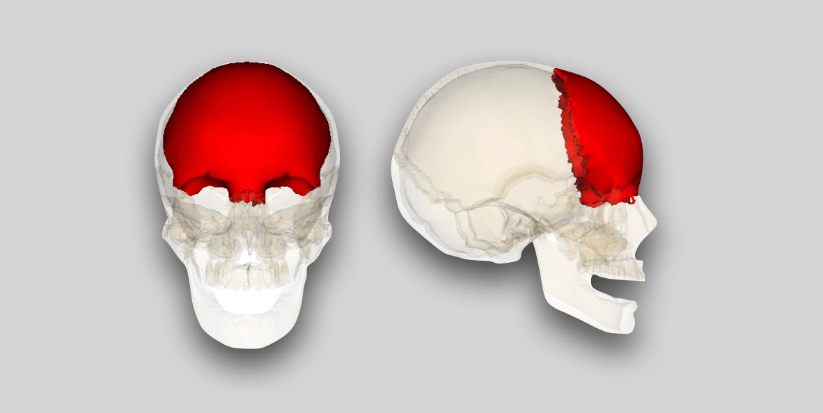
Frontal bone is shell-shaped unpaired bone located in the forehead region. It forms the forehead, most of the roof of orbit and floor of anterior cranial fossa.
It consists of three parts: Squamous part, Orbital plates and Nasal part. The junction of squamous part and orbital plate forms the supraorbital margin.
 Source: Sobotta's Atlas and Text-book of Human Anatomy 1909, Author: Dr. Johannes Sobotta
Squamous Part
The squamous part presents two surfaces and two borders, and encloses a pair of frontal air sinuses.
External/Outer Surface
It is smooth and presents the following features:
- Supraorbital foramen: It is present along the supraorbital margin, at the junction of medial 1/3rd and lateral 2/3rd. It transmits the supraorbital nerve and vessels.
- Superciliary arch: Curved elevation above each supraorbital margin.
- Glabella: Median round prominence between the medial ends of the two superciliary arches.
- Frontal tuber/tuberosity/eminence: Round elevation above the superciliary arch, on each side.
- Zygomatic processes: These extends downward and laterally, on each side, from the lateral end of supraorbital margin. These articulate with the frontal process of zygomatic bone, forming the frontozygomatic suture.
- Temporal line: It curves upward and backwards from the posterior margin of each zygomatic process.
- Temporal surface: It is the area below the temporal line and behind the zygomatic process.
- Metopic suture: Fibrous joint that divided the two halves of the frontal bone in infants. It is the first suture to close physiologically, as early as 3 months (complete fusion by 8 months).
Internal/Inner Surface
It is deeply concave and presents:
- Anterior portion of sagittal sulcus: Sagittal sulcus is a shallow depression/groove found along the midline on the internal surface of the frontal, parietal and occipital bones. It houses/lodges the superior sagittal sinus and its margin provide attachment sites for falx cerebri.
- Frontal crest: Bony ridge located in the midline of the internal surface, formed by the convergence of the edges of sagittal sulcus.
 Source: Sobotta's Atlas and Text-book of Human Anatomy 1909, Author: Dr. Johannes Sobotta
Borders
- Upper/Parietal border.
- Lower/Orbital border.
Frontal Air Sinus
- These are paired triangular or trapezoidal-shaped air-filled cavities that lies between the outer and inner tables of frontal bone, above the supraorbital margin.
- It is larger in men than in women and is responsible for the prominent frontal bossing seen in many men.
- It drains into the anterior portion of the middle meatus through the frontonasal duct.
Orbital Plates
These are triangular, curved plates of bone extending horizontally backwards from the supraorbital margin, and are separated by a wide gap, called the ethmoidal notch.
Inferior/Orbital surface
It is smooth and concave, and presents:
- Lacrimal fossa (anterolaterally).
- Trochlear spine (anteromedially).
Superior/Cerebral surface
- It is convex and forms most of the floor of anterior cranial fossa.
- It presents depressions for the convolutions of frontal lobes of the brain.
Ethmoidal Notch
- U-shaped notch that separates the two orbital plates.
- It is quadrilateral and occupied by cribriform plate of ethmoid bone.
- Each side of notch presents small air spaces (half-cells), that articulates with the labyrinth of ethmoid to complete ethmoidal air sinuses/cells.
- The margins are transversed by grooves that form anterior and posterior ethmoidal canals.
 Source: Sobotta's Atlas and Text-book of Human Anatomy 1909, Author: Dr. Johannes Sobotta
Nasal Part
It is part of frontal bone projecting downwards between the two supraorbital margins (right and left). It presents a nasal notch.
Articulation of Nasal Notch
- Inferiorly: Nasal bones, on each side of median plane.
- Laterally: Frontal process of maxilla and lacrimal bone.
Anatomical Position
In the anatomical position,
- Squamous part is vertical and convex forwards.
- The two orbital plates are horizontal, projecting backwards.
- Nasal part is directed forwards and downwards.
 Source: Wikimedia Commons. Author: en:Anatomography
Applied Anatomy
-
Trigonocephaly: A type of craniosynostosis characterised by triangular, or wedge-shaped forehead, resulting from the premature fusion and ossification of the metopic suture.
-
The squamous part of frontal bone is prone to fracture. In neonates and
infants, a depressed fracture (a dimple in the bone) occurs, while adult skull shows fissured fracture, i.e., the depressed area with an irregular line of fracture at periphery.
-
The frontal sinus can be a source of orbital cellulitis, but it more commonly affects the orbit with slowly progressive growth of a frontal sinus mucocele (due to obstruction of the sinus drainage).
References
- BD Chaurasia's Human Anatomy Volume 3 (Head & Neck), Eighth edition, CBS Publishers and Distributors Pvt Ltd
- Textbook of Anatomy Head, Neck, and Brain (Volume III), Vishram Singh
- van der Meulen J. Metopic synostosis. Childs Nerv Syst. 2012 Sep;28(9):1359-67. doi: 10.1007/s00381-012-1803-z. Epub 2012 Aug 8. PMID: 22872249; PMCID: PMC3413823.
- Cover Image Credit: Source - Anatomography[3] website maintained by Life Science Databases(LSDB). This file is made available under the Creative Commons Attribution-Share Alike 2.1 Japan license.
*This article is an excerpt from the above mentioned sources and Medical Sutras does not make any ownership or affiliation claims.