The permanent mandibular lateral or second incisors are the second mandibular tooth from the median line. It is situated distal to the mandibular central incisors and mesial to the mandibular canine.
It resembles mandibular central incisor very closely, as the two teeth operate as a team and their functional form is related. However, it is somewhat larger than mandibular central incisor.
Chronology
- First evidence of calcification: 3-4 months.
- Crown completion: 4-5 years.
- Eruption: 7-8 years.
- Root completion: 10 years.
Dimensions
- Overall length: 23.5 mm.
- Crown length: 9.5 mm.
- Root length: 14.0 mm.
- Mesiodistal diameter of crown: 5.5 mm.
- Mesiodistal diameter of crown at cervix: 4.0 mm.
- Labiolingual diameter of crown: 6.5 mm.
- Labiolingual diameter of crown at cervix: 5.8 mm.
- Curvature of cervical line (Mesial): 3.0 mm.
- Curvature of cervical line (Distal): 2.0 mm.
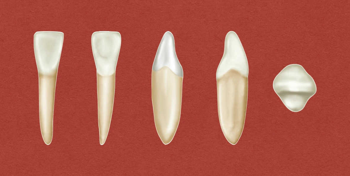
Crown Morphology
Labial & Lingual Aspects
- These are similar to that of mandibular central incisor, except for the mesiodistal diameter, which is about 1 mm more (added to the distal half).
- In some Mongoloid groups, the lingual aspect presents a deep but short cervicoincisal groove, which is vulnerable to dental caries.
Mesial & Distal Aspects
- No marked difference is evident as compared to the mesial and distal surfaces of lower central incisor, except for the size.
- Mesial side is often longer than the distal side, resulting in the sloping of the incisal ridge downward in distal direction.
- Distal contact area is more cervical than the mesial contact area, as it has to form proper contact with the mesial side of mandibular canine.
- The distal surface presents a tendency for deeper concavity immediately above the cervical line.
Incisal Aspect
- Presents a characteristic feature to identify the tooth and distinguish it from central incisor: Incisal edge is not at approximate right angle to the line bisecting crown and root labiolingually, as found in central incisor.
- Incisal edge follows the curvature of lower arch, making the crown appear slightly twisted on its root base.
- The labiolingual root axes of mandibular central and lateral incisors are almost parallel in the alveolar process.
Root Morphology
- The root form is similar to that of the central incisor. However, it may be considerably longer than that of central incisor.
- Developmental depressions are present on both mesial and distal side.
References
- Wheeler's Dental Anatomy, Physiology and Occlusion(2019), Stanley J. Nelson DDS MS, Elsevier.
*This article is excerpt from the above mentioned book and Medical Sutras does not make any ownership and affiliation claims.