The term molar comes from the Latin word "Molaris dens" meaning grinding tooth. It is derived from the word "mola" which refers to millstone (a pair of large stones used for grinding food).
Function
- As the name implies, the function of molars is to crush and grind food into small pieces that can be swallowed easily.
- Also, they help maintain vertical height of the face and supports facial muscles.
Position
- They are the 6th teeth from midline, present posterior to Maxillary 2nd Premolar and anterior to Maxillary 2nd Molar.
- The normal location of permanent maxillary first molar is at the center of fully developed adult jaw anteroposteriorly. Hence, they are also considered as cornerstones of the dental arches.
Tooth Numbering
- FDI system: 16 (Permanent Maxillary Right 1st Molar), 26 (Permanent Maxillary Left 1st Molar).
- Universal system: 3 (Permanent Maxillary Right 1st Molar), 14 (Permanent Maxillary Left 1st Molar).
- Zsigmondy-Palmer system: 6| (Permanent Maxillary Right 1st Molar), |6 (Permanent Maxillary Left 1st Molar).
Chronology
- First evidence of calcification: At birth
- Crown completion: 3-4 years
- Eruption: 6 years
- Root completion: 9-10 years
Dimensions
- Overall length: 19.5mm
- Crown length: 7.5mm
- Root length: 12mm (Buccal), 13mm (Palatal)
- Mesiodistal diameter of crown: 10mm
- Mesiodistal diameter of crown at cervix: 8.0mm
- Labiolingual diameter of crown: 11mm
- Labiolingual diameter of crown at cervix: 10mm
Surfaces, Lines Angles, Point Angles
- Surfaces (5): Buccal, Lingual, Mesial, Distal, Occlusal
- Line Angles (8): Mesiobuccal, Distobuccal, Mesiolingual, Distolingual, Mesioocclusal, Buccoocclusal, Distoocclusal, Linguoocclusal
- Point Angles (4): Mesiobuccoocclusal, Distobuccoocclusal, Distolinguoocclusal, Mesiolinguoocclusal
Tooth Morphology
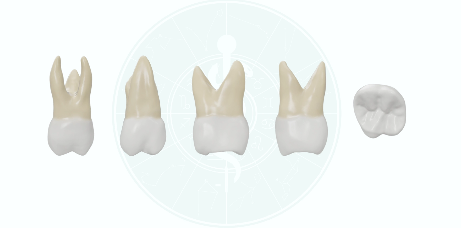
Buccal Aspect
- Crown outline/shape: Trapezoidal (shorter of uneven sides toward the cervix).
- Mesial outline: Almost straight near the cervical region and curves occlusally as it reaches the mesial contact area and joins the mesial slope of the MB cusp. The mesial contact area is at the occlusal third of the crown i.e., two-thirds the distance from cervical line to the tip of MB cusp.
- Distal outline: More convex from the cervix to the point it joins the occlusal outline or distal slope of DB cusp. The distal contact area is at the middle third of the crown.
- Cervical outline: Irregular and curves slightly in apical direction.
- Occlusal outline: Formed by the MB and DB cusp tips and their slopes.
- Cusps: MB cusp is broader than DB cusp and the cusp slopes meet at an obtuse angle. On the other hand, the mesial and distal slopes of DB cusp meet at approximately right angle, thus, making the DB cusp appear sharper than MB cusp.
- Buccal Developmental Groove: Divides the occlusal outline or MB and DB cusps at the centre, and terminates at a point approximately half the distance from occlusal outline to the cervical line. The groove is not deep at any point, however, it becomes more shallow towards its end and gradually fades out.
Lingual Aspect
- Crown outline: Trapezoidal shape (shorter of uneven sides towards the cervix).
- Mesial outline: Follows a straight path from cervix and curves as it joins the mesial slope of ML cusp.
- Distal outline: Convex and confluent with the DL cusp, forms a semicircular arc.
- Cervical outline: Almost straight on the lingual aspect.
- Occlusal outline: Formed by the ML and DL cusp tips and their slopes.
- Cusps: ML cusp is larger and is the longest cusp of Permanent Maxillary 1st Molar. Its mesiodistal width is approximately three-fifth the mesiodistal width of crown, while DL cusp makes up the remaining two-fifth. The mesial and distal slopes of ML cusp meets at an obtuse angle. On the other hand, the DL cusp is spheroidal in shape and it is difficult to describe any angulation on its mesial and distal slopes.
- Lingual Developmental Groove: Starts approximately in the centre of the lingual surface and curves distally as it crosses between the lingual cusps onto the occlusal surface.
- Cusp of Carabelli (5th cusp): It is the most important feature of lingual surface of Maxillary 1st Molar. It appears attached to the ML surface of the ML cusp. The cusp ridge of 5th cusp is approximately 2mm cervical to the ridge of ML cusp and is usually separated by a groove.
Mesial Aspect
- Crown outline: Trapezoidal shape (shorter of uneven side occlusally).
- Buccal outline: Convex in the cervical region, then becomes less convex or straight as it joins the MB cusp. he crest of curvature is in the cervical thid.
- Lingual outline: Forms a more convex arc from cervix to ML cusp. The crest of curvature is near the middle third of the crown.
- Cervical outline: Irregular and curves occlusally upto 1mm.
- Occlusal outline: Formed by MMR and the MB and ML cusp ridges.
- Cusps: The MB and ML cusp are within the confines of the root trunk. The MB cusp tip is on line with the long axis of the lingual root, while the ML cusp tip is on line with the buccal outline of the MB root.
Distal Aspect
- The crown is trapezoidal in shape and appears similar to the mesial aspect.
- Variations on the distal aspect include:
- More of the buccal surface can be seen from the distal aspect, because of the tendency of crown to taper distally.
- DMR dips sharply in cervical direction. It is shorter and at a lower level than the MMR, hence, some part of the occlusal surface and triangular ridges can be seen.
- Cervical outline is almost straight without much curvature.
Occlusal Aspect
-
Shape: Rhomboidal (Acute angles - MB and DL; Obtuse angles - DB and ML)
-
Dimensions
- Buccolingual dimension of crown (11mm) is greater than the MD dimension (10mm).
- Buccolingual measurement on the mesial side (mesial to buccal and lingual developmental grooves) is greater than the BL measurement on the distal side. This is because the crown tapers distally.
- Mesiodistal measurement of crown lingual to the contact areas is greater than MD measurement bucally, i.e., the crown does not show lingual convergence as seen in most permanent teeth.
-
Elevations
- Cusps: There are 4 major cusps (MB, ML, DB, DL) and a supplemental fifth cusp (Cusp of Carabelli). The ML cusp is the largest, followed by MB, DL, DB and 5th cusp.
- Cusp Ridges: Each cusp have mesial and distal cusp ridges and a triangular ridge sloping towards the centre of the occlusal surface.
- Oblique Ridge: It is formed by the unoin of triangular ridge of distobuccal cusp and distal ridge of ML cusp, and crosses the occlusal surface obliquely (hence the name).
- Marginal Ridges: The tooth have prominent mesial and distal marginal ridges that forms the mesial and distal boundary of the occlusal surface respectively.
-
Maxillary Molar Primary Cusp Triangle
- It is an important feature characteristic of all maxillary molars.
- Formed by the triangular arrangement of the three primary cusps (ML, MB, DB cusps), alongwith the mesial marginal ridge and oblique ridge.
-
Depressions
- Major Fossae (2): Central fossa (triangualar in shape, present mesial to oblique ridge); Distal fossa (roughly linear, present distal to oblibue ridge).
- Minor Fossae (2): Mesial triangular fossa (distal to MMR); Distal triangular fossa (mesial to DMR).
- Pits (3): Central pit (pin-point depression in the central fossa); Mesial pit (at the apex of MTF); Distal pit (at the apex of DTF).
- Developmental Grooves (5):
- Buccal DG: Runs buccally from the CP and continues onto the buccal surface separating the two buccal cusps.
- Central DG: Runs in mesial direction and ends at the apex of MTF, separates the triangular ridges of MB and ML cusps.
- Transverse groove of oblique ridge: Runs in distolingual direction from CP, crossing the oblique ridge to reach distal fossa.
- Distal oblique groove: Runs in oblique direction parallel to oblique ridge and joins the lingual DG present on the lingual surface; separates DL cusp from the rest of occlusal surface.
- Fifth cusp groove: Separates the fifth cusp from ML cusp.
- Supplemental Grooves: Several supplemental grooves are present at the apices of mesial and distal triangular fossae.
References
- Wheeler's Dental Anatomy, Physiology and Occlusion, Stanley J. Nelson DDS MS, Elsevier
- Textbook Of Dental Anatomy, Physiology & Occlusion, Rashmi GS Phulari, Jaypee Brothers Medical Publishers Pvt Ltd
*This article is excerpt from the above mentioned book and Medical Sutras does not make any ownership and affiliation claims.